Key points
- A 2015 meta-analysis found mean adult effective doses of 84 microsieverts for small, 177 for medium and 212 for large fields of view, with large differences between machines.
- Two panoramic machines measured 14.2 and 24.3 microsieverts in one phantom study, so a CBCT scan usually delivers more than a panoramic X-ray.
- The field of view and exposure settings drive most of the difference, which is why guidelines ask for a reason for every scan.
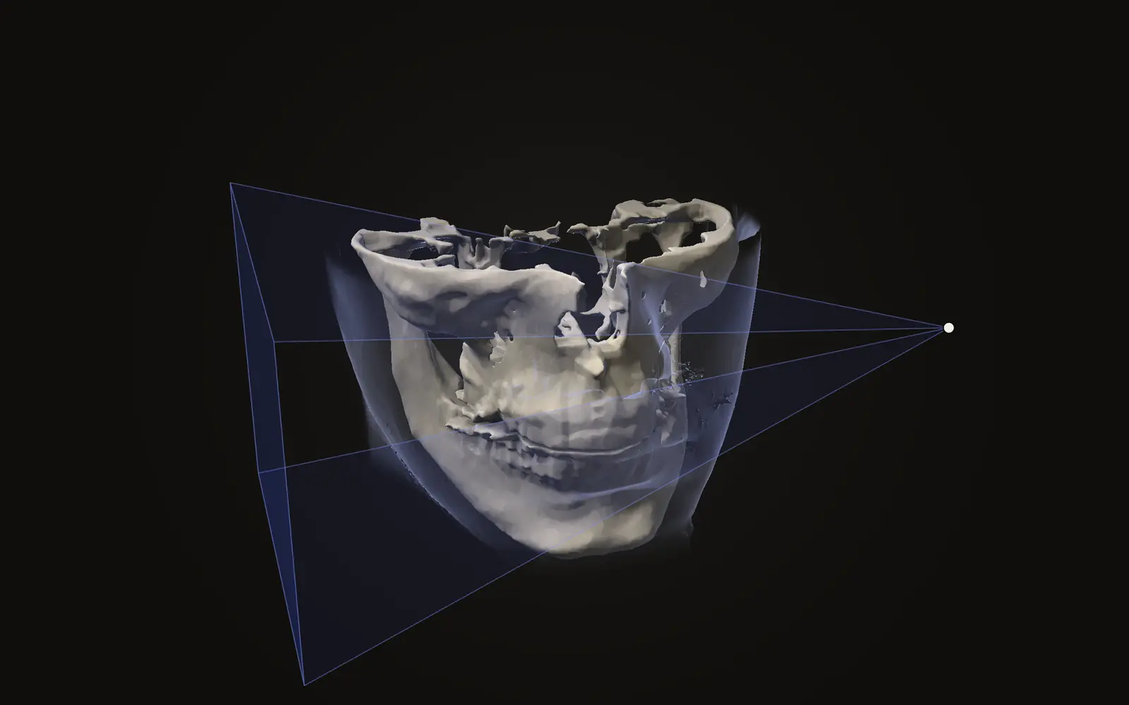
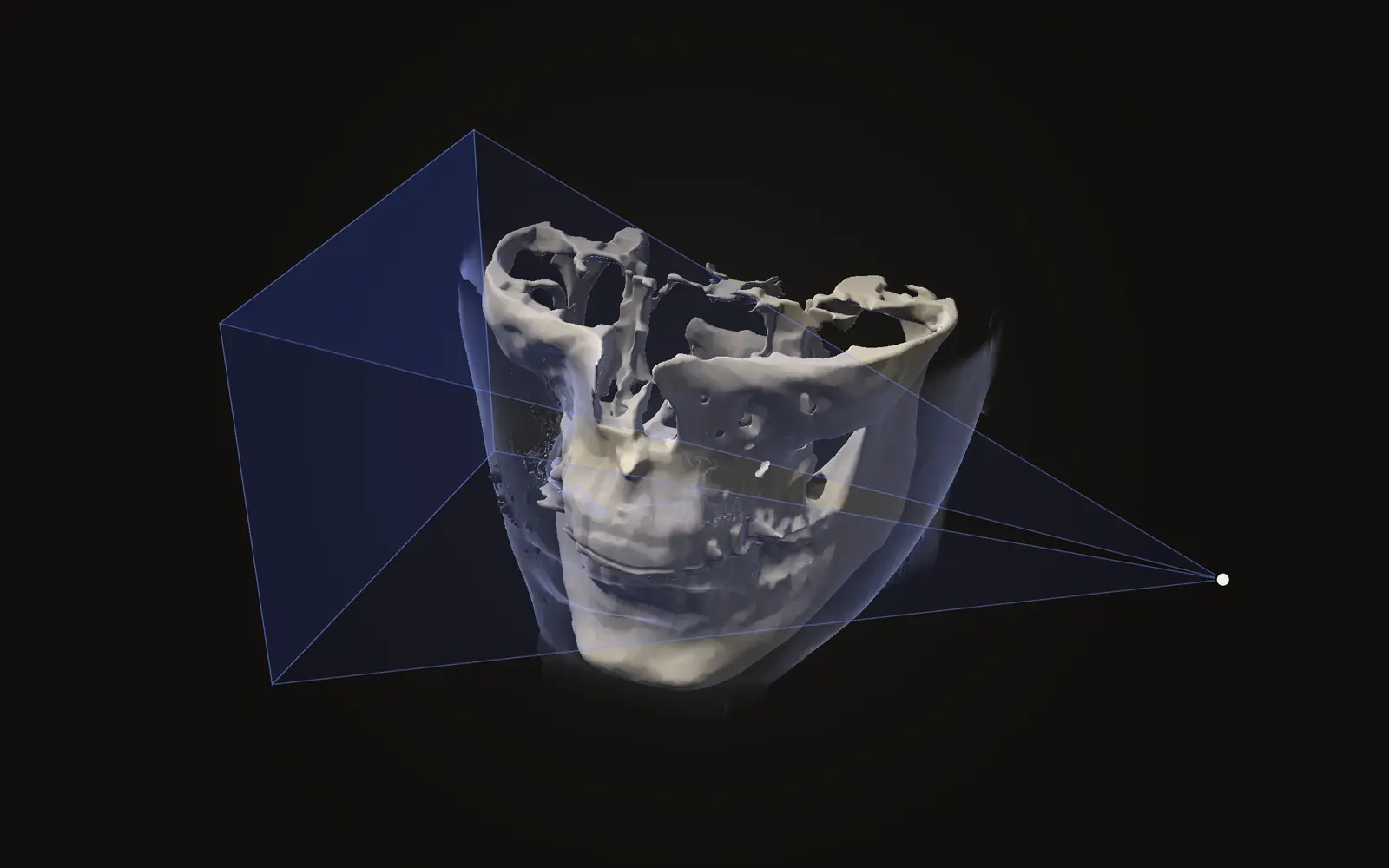
The radiation dose from a CBCT scan varies widely. Published measurements put that range in context alongside panoramic X-rays, medical CT and everyday background radiation.
What the number means#
Doses in dental imaging are quoted as effective dose, in microsieverts (µSv). Effective dose adds up the dose to each organ in the beam, weighted by how sensitive that organ is, so that different examinations can be compared. It is the preferred way to compare doses between types of exposure.[1]
The weights come from the 2007 recommendations of the International Commission on Radiological Protection (ICRP). Those recommendations include the salivary glands and the lining of the mouth, which earlier ones did not. Re-calculated that way, dental doses came out 23 to 224 percent higher than under the 1990 weights in one study.[2] So an older figure and a newer one for the same scan can differ because of the method, not the machine. Check which recommendation a number uses before you compare.
What studies measured for CBCT#
The largest overview is a 2015 meta-analysis. After screening 519 and 743 search results, the authors kept 20 publications and added measurements on nine more units.[3]
84 µSv
mean adult dose, small field of view[3]
177 µSv
mean adult dose, medium field of view[3]
212 µSv
mean adult dose, large field of view[3]
Those are means for standard protocols. The full spread was far larger: adult doses for any protocol ranged from 46 to 1073 µSv with large fields of view, 9 to 560 µSv with medium fields and 5 to 652 µSv with small fields. For children, the mean was 175 µSv for large and medium fields combined and 103 µSv for small fields.[3] The authors say the large exposure range makes CBCT doses hard to generalise.
Different machines account for much of the spread. A separate study measured 14 devices in head phantoms and found effective doses between 19 and 368 µSv, a 20-fold range.[4] Most of the dose went to three groups of tissue: the remaining tissues (37 percent), the salivary glands (24 percent) and the thyroid (21 percent). The authors conclude that small-, medium- and large-field scanners need to be treated separately, because the dose is strongly tied to the field of view, and that dose should always be weighed against the image quality the question needs.[4]
Next to a panoramic X-ray and a medical CT#
A panoramic radiograph is a single flat image. In one phantom study, two panoramic machines gave 14.2 and 24.3 µSv. A lateral skull X-ray (cephalogram) gave 5.6 µSv, and four bitewing images gave 5.0 µSv.[1]
For medical CT, the same group measured one 64-slice unit alongside eight CBCT units. For a similar field of view, the CT gave 860 µSv, while medium-field CBCT units gave 69 to 560 µSv and large-field units 68 to 1073 µSv.[2] The authors recommend dental CBCT as the lower-dose choice over a medical CT for common jaw imaging tasks. Keep the limits in mind: that is one CT unit measured in 2008, and the large-field CBCT range reaches above it. It does not describe every CT scanner or protocol.
| Examination | Effective dose (µSv) | Source |
|---|---|---|
| Four bitewing images | 5.0 | [1] |
| Lateral cephalogram | 5.6 | [1] |
| Panoramic X-ray, two machines | 14.2 and 24.3 | [1] |
| CBCT, small field, mean | 84 | [3] |
| CBCT, medium field, mean | 177 | [3] |
| CBCT, large field, mean | 212 | [3] |
| 64-slice CT, one unit | 860 | [2] |
Next to background radiation#
Everyone receives radiation from the ground, the air, food and space. The worldwide average is about 2.4 millisieverts a year, which is 2400 µSv.[5] Spread over a year, that is roughly 6.6 µSv a day. By that arithmetic (ours, not from a study), a mean large-field CBCT scan of 212 µSv equals about 32 days of natural background, and a mean small-field scan of 84 µSv about 13 days. Background varies by place, and the same fact sheet gives 1.8 mSv a year as the Canadian average.[5]
Why guidelines ask for a reason#
Because dose varies this much, the European Academy of Dental and Maxillofacial Radiology agreed a set of 20 basic principles for dental CBCT, built on European radiation-protection directives and tested with its members in a consensus process.[6] A study of everyday dental exposures concluded that, even though radiographs are an indispensable diagnostic tool, the doses are high enough to justify looking for ways to reduce them, such as digital receptors and selecting the right examination for the question.[1]
Every examination should have a reason, using the smallest field that answers the question.
What these studies do not tell you#
Most dose figures come from phantoms, which are models of a head built to stand in for a patient. They show what a protocol deposits in the organs, not what happened to you. The studies here are from 2008 to 2015, and machines and protocols have changed since. Newer low-dose protocols exist on many units, which the 2015 meta-analysis notes extend the range of doses further.[3]
How much radiation is in a dental CBCT scan?
Studies report a wide range. A 2015 meta-analysis found mean adult effective doses of 84 microsieverts for small fields of view, 177 for medium and 212 for large, with large differences between machines.
Is a CBCT scan more radiation than a panoramic X-ray?
Usually yes. Two panoramic machines measured 14.2 and 24.3 microsieverts in one study, while the mean for a large-field CBCT scan was 212 microsieverts in another.
Is CBCT less radiation than a medical CT?
In a 2008 phantom study, a similar-sized medical CT scan gave 860 microsieverts against 69 to 560 for medium-field CBCT units. That was one CT unit, so it is not a general figure for CT.
Why does the dose differ so much between scans?
Mainly the size of the field of view, the exposure settings, and where the beam sits relative to sensitive organs such as the salivary glands and thyroid.
Sources
- Ludlow JB, Davies-Ludlow LE, White SC (2008). Patient risk related to common dental radiographic examinations: the impact of 2007 International Commission on Radiological Protection recommendations regarding dose calculation. J Am Dent Assoc, 139: 1237-43.DOIPubMed
- Ludlow JB, Ivanovic M (2008). Comparative dosimetry of dental CBCT devices and 64-slice CT for oral and maxillofacial radiology. Oral Surg Oral Med Oral Pathol Oral Radiol Endod, 106: 106-14. Eight CBCT units and one 64-slice CT unit, measured in a phantom.DOIPubMed
- Ludlow JB, Timothy R, Walker C, et al. (2015). Effective dose of dental CBCT, a meta analysis of published data and additional data for nine CBCT units. Dentomaxillofac Radiol, 44: 20140197. Meta-analysis of 20 publications plus new measurements on nine units.DOIPubMed
- Pauwels R, Beinsberger J, Collaert B, et al. (2012). Effective dose range for dental cone beam computed tomography scanners. Eur J Radiol, 81: 267-71. Phantom measurements on 14 devices.DOIPubMed
- Canadian Nuclear Safety Commission (2020). Natural background radiation (fact sheet). Canadian Nuclear Safety Commission. States the worldwide average natural background dose as 2.4 mSv a year.Link
- Horner K, Islam M, Flygare L, et al. (2009). Basic principles for use of dental cone beam computed tomography: consensus guidelines of the European Academy of Dental and Maxillofacial Radiology. Dentomaxillofac Radiol, 38: 187-95.DOIPubMed
Terms used
- Effective dose
- A single number in sieverts that adds up the dose to each organ, weighted by how sensitive that organ is to radiation. It lets you compare different examinations.
- Microsievert
- One millionth of a sievert (µSv). Dental imaging doses are usually quoted in microsieverts.
- Field of view
- The size of the region a scan covers. A small field of view shows a few teeth; a large one shows the whole head.
- Panoramic radiograph
- A single flat dental X-ray that shows both jaws and all teeth in one image.
This is general information, not medical advice. Jaw Viewer is not a medical device. Talk to your surgeon, orthodontist or radiologist about your own case.