See inside your own jaw scan.
Open the CT or CBCT files from your clinic. Look around in 3D, take measurements or compare two dates. Your files stay on your computer.
Free for personal use. No account, nothing to install.
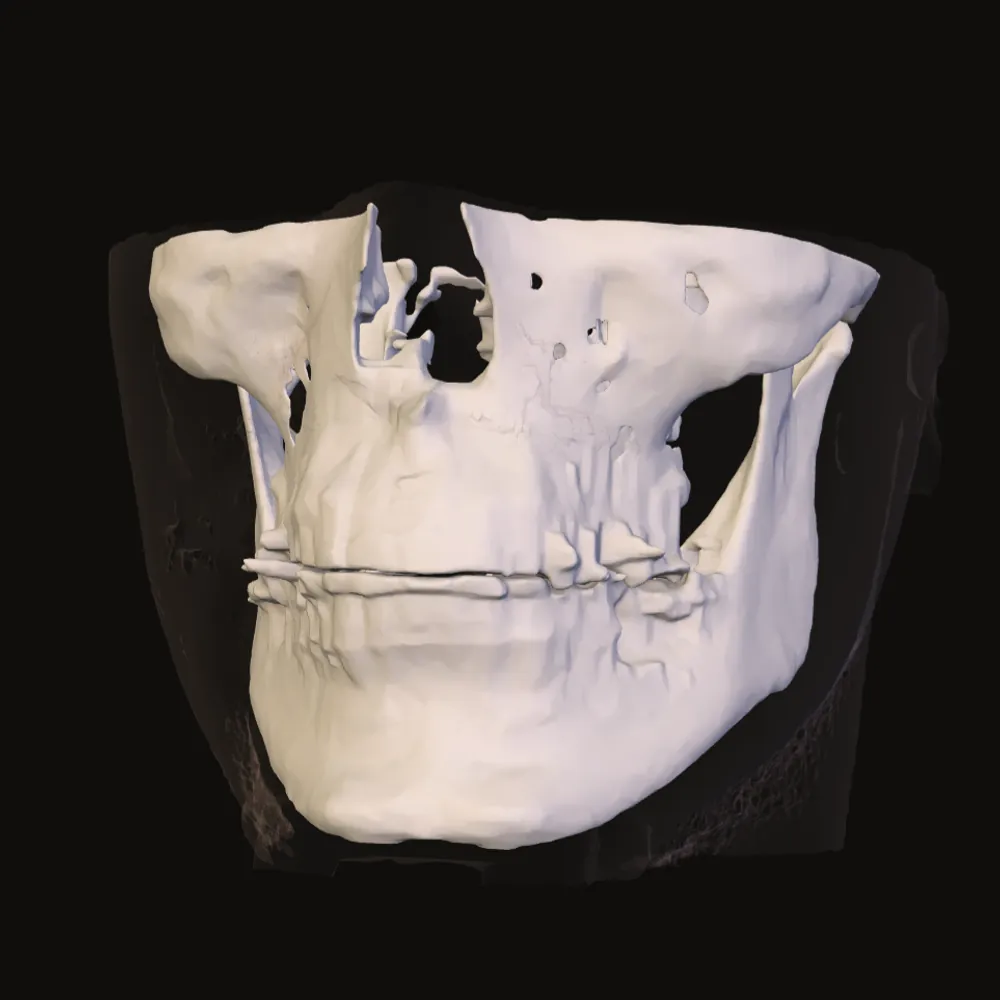
Open the CT or CBCT files from your clinic. Look around in 3D, take measurements or compare two dates. Your files stay on your computer.
Free for personal use. No account, nothing to install.
After my jaw operation I had two scans on two discs and no way to look at them properly, let alone next to each other.
So I built a viewer that runs in the browser and keeps the scans on my computer.
Drag to turn the scan. Fade the skin, look at the teeth, or cut away the bone that is in the way.
You work from the files your clinic gave you. They stay unchanged.
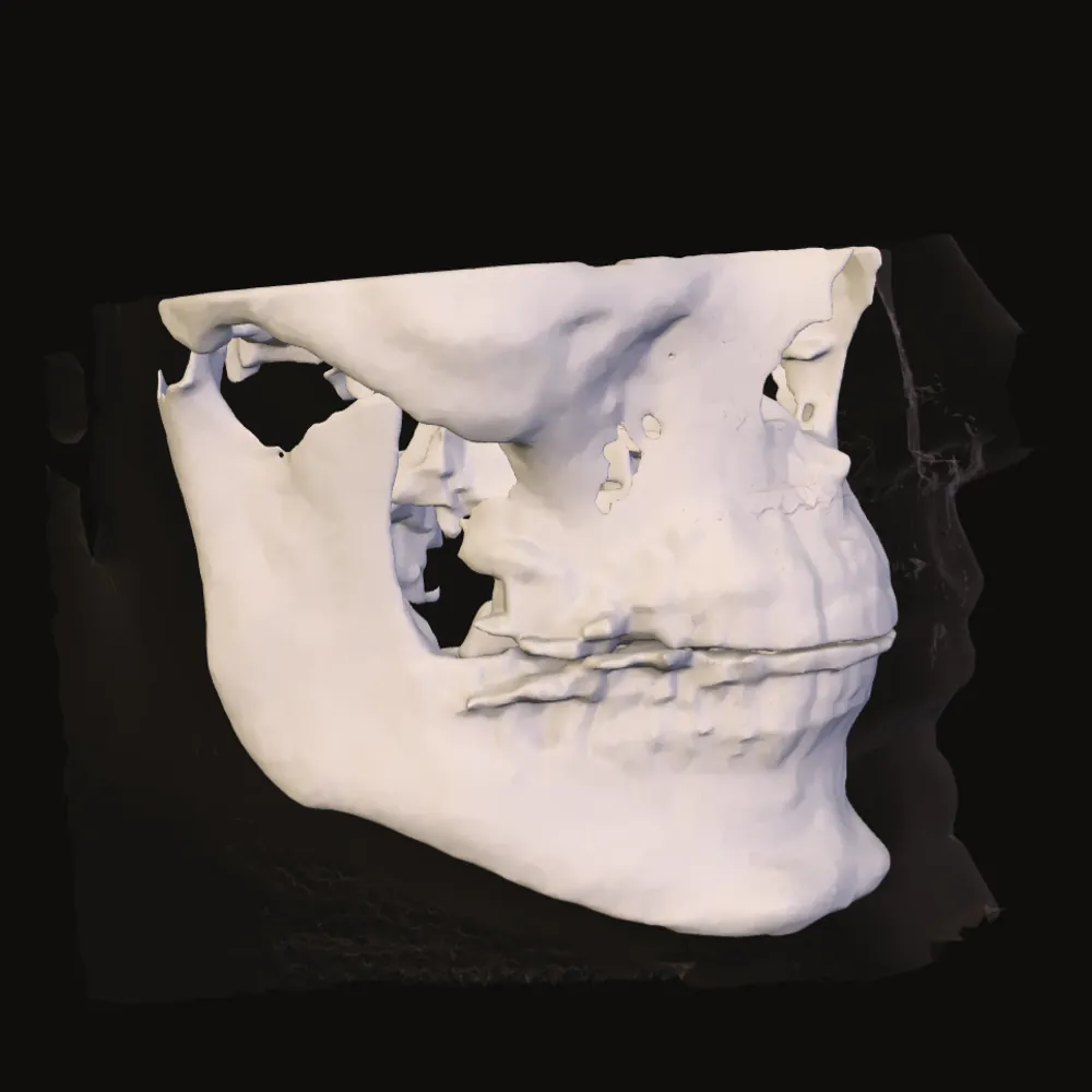
Show the skin surface the scanner recorded and turn it to see the shape of the lower face.
This sample ends below the nose and at the neck. The surface shows exactly what was scanned, cut-off edges included.
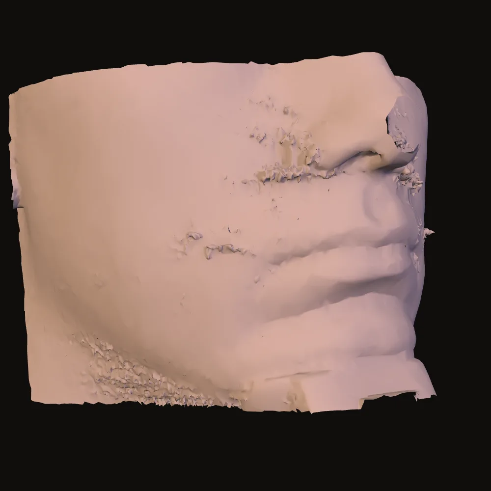
Fade the skin to see the teeth and jaws through the outline of the face.
Skin and bone come from the same scan, so you can switch between them without losing your place.
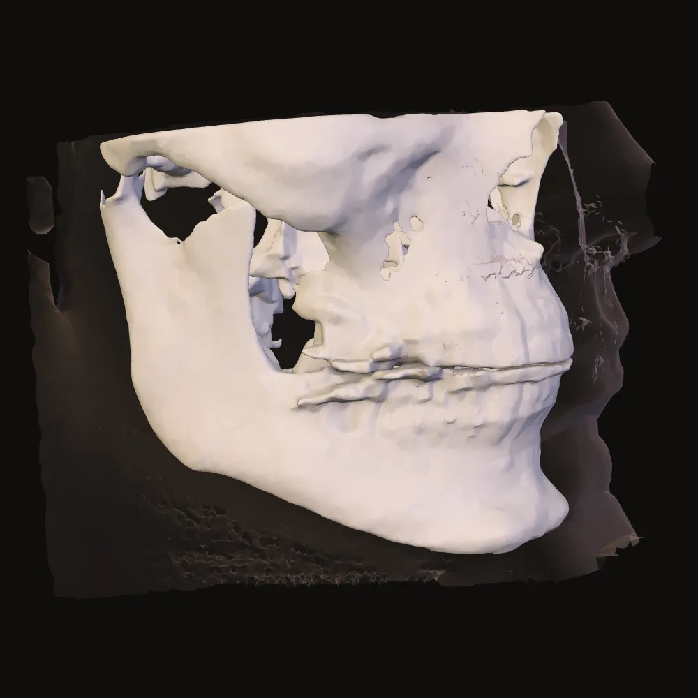
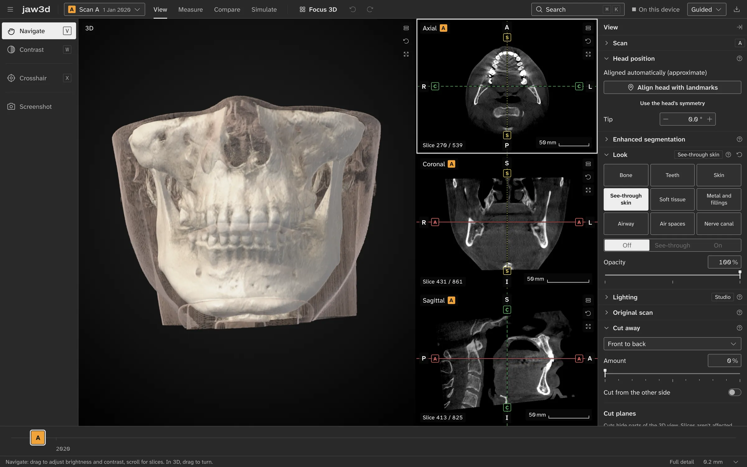
The 3D view gives you the shape. Slices let you look through it, from above, from the front and from the side.
Click a point in one view to find the same place in the others.
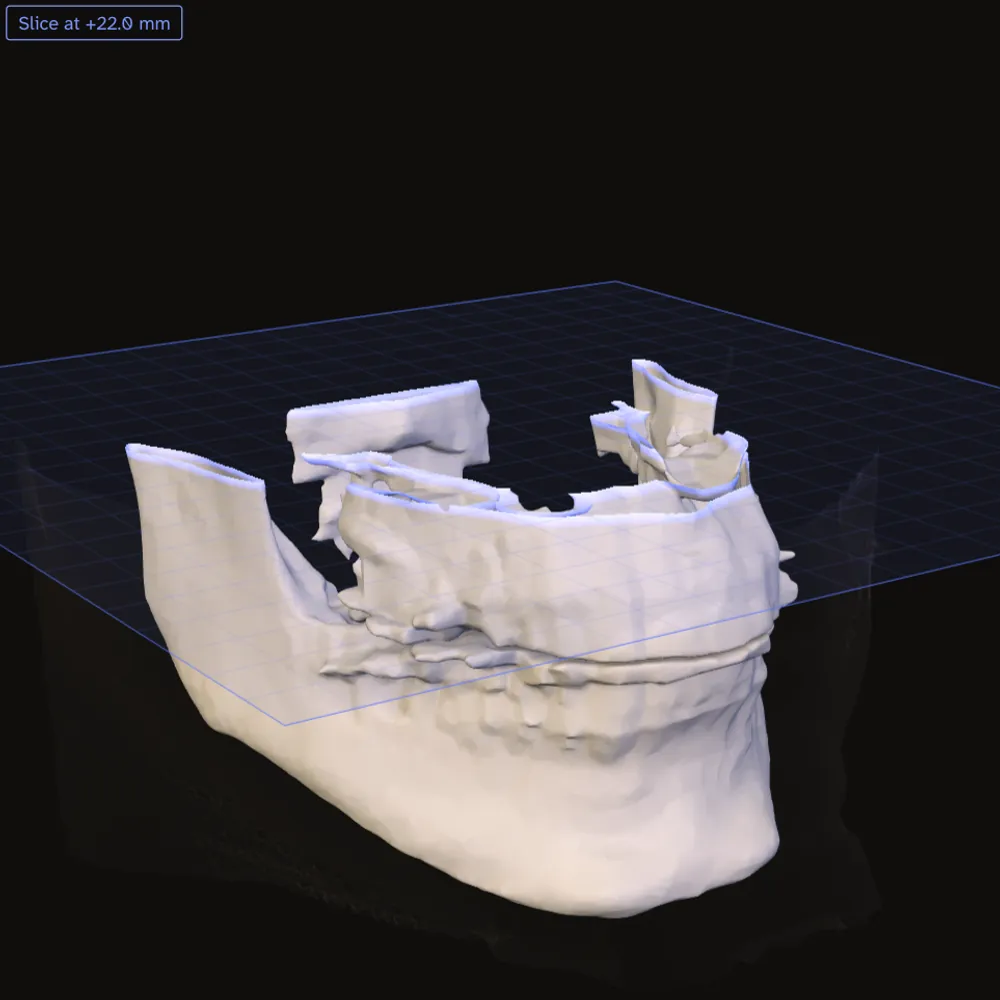
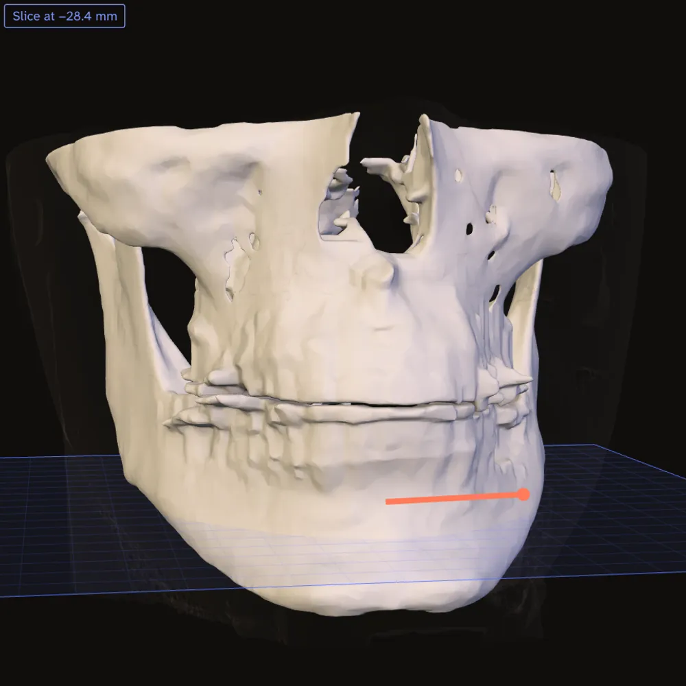
Two points make a distance, three an angle. The same line shows in the slices and on the bone.
The voxel size beside each value helps you judge its precision. Landmarks, the airway and the sinuses have their own tools.
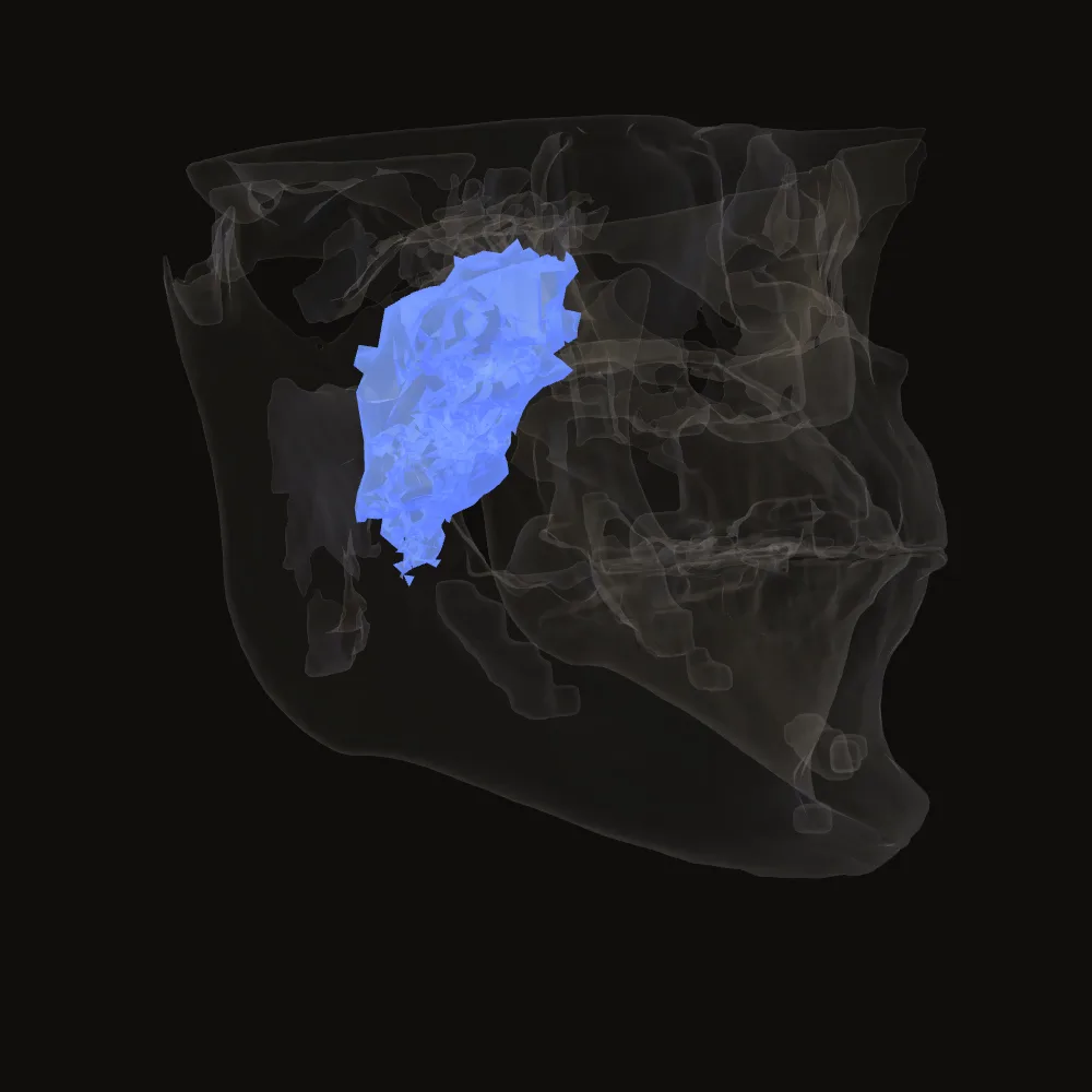
Fade the bone and the part of the airway caught in the scan appears behind the mouth.
In Measure you check its borders in the slices, get a volume and see where it is narrowest.
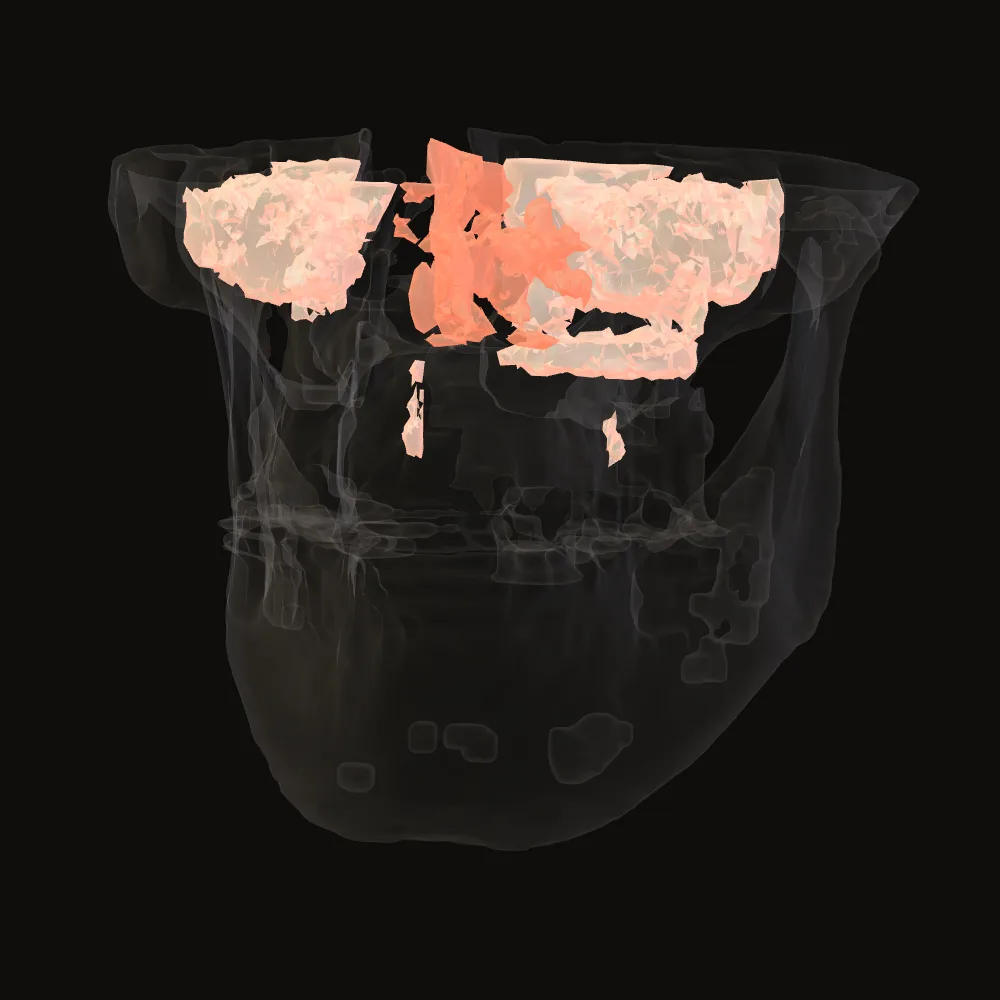
See the nasal cavity and both maxillary sinuses, with a volume in millilitres for each.
The number belongs to the region the tool selected. Check its borders in the slices before you read anything into it.
Before you compare angles, check which way the head was held in the scanner. Landmarks you place tell the viewer which way is up and which way is forward.
Check the reference planes against your own anatomy before relying on them.
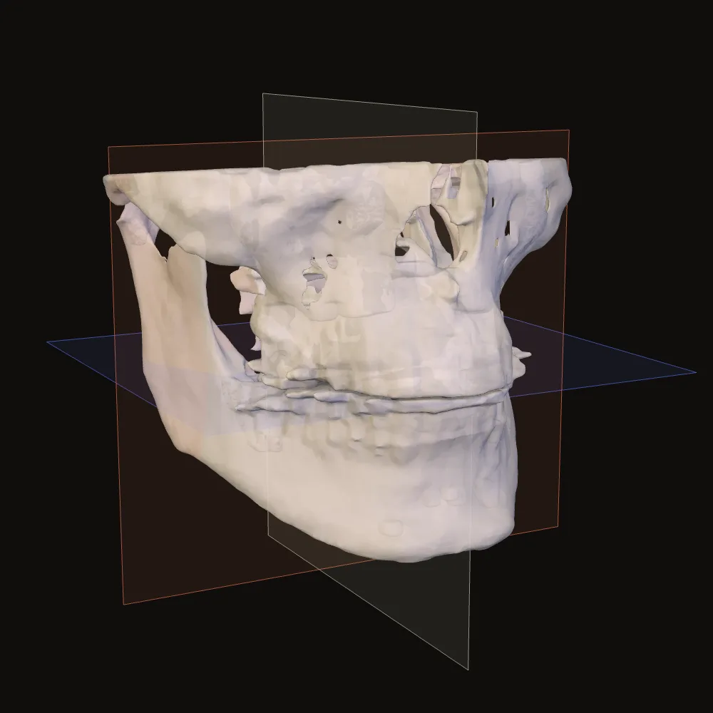
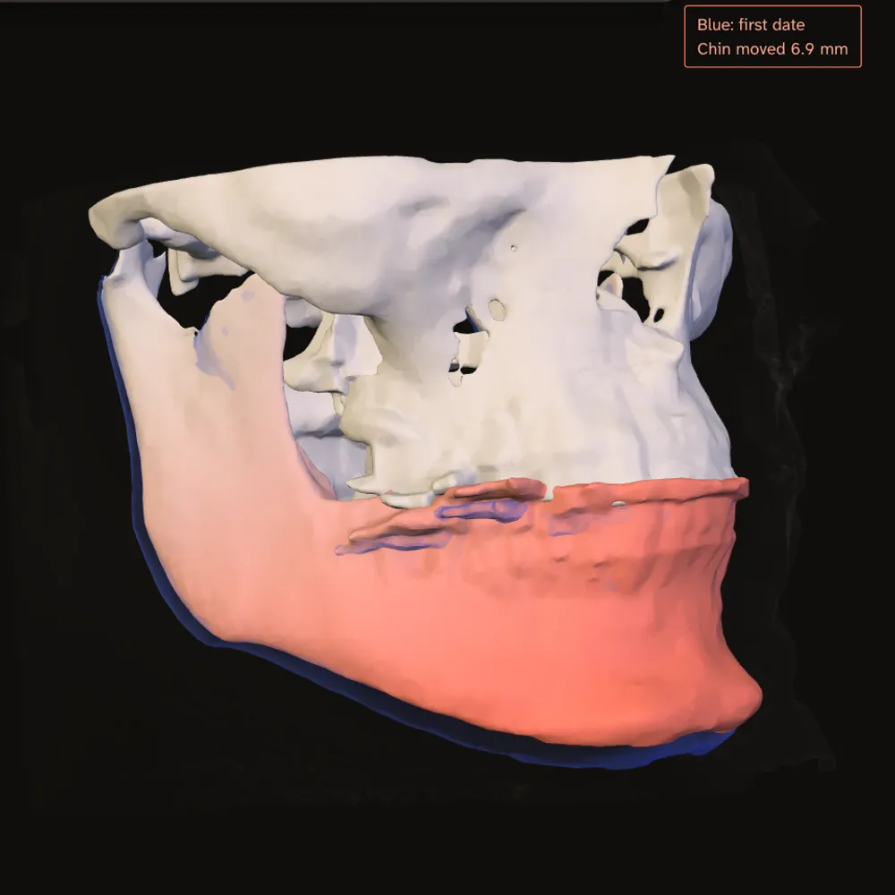
Line up two scans on the skull base. Compare them side by side or swipe between dates; a distance map shows how far apart their bone surfaces are.
The blue ghost marks the first date. Here, the chin has the largest change.
Made with one public sample moved by hand to stand in for a second date. Your own comparison needs two scans.
Drag to turn the sample skull. Try slicing from the top, measuring the jaw or comparing two dates. When you open your own files, three slice views sit beside the 3D view.
Drag to turn the skull. With the view focused, the arrow keys turn it too and Home puts it back. The sample scan ends below the eyes, so the top of the skull fades out.
The second date is the same public sample with its lower jaw moved by hand. Your own comparison needs two scans.
Find the same point in 3D and in the slices. Start in View, then choose Measure or Compare to look closer.
A folder from a disc, a zip download or the DICOM files themselves all work. A computer gives you the most room.
Help opening your first scanYour browser loads the viewer from this site. You do not need an account.
Use Choose folder or Choose files, or drop them into the viewer window.
Open the scan card, turn the 3D view and scroll through the slices.
Your browser reads the files, draws the views and runs the measurements and comparisons. Your scan is never uploaded.
What is downloaded, stored and savedThe site sends your browser the viewer. Your scan files are not sent back.
You choose when to save a project and whether to include the scan images. Anonymous usage counts in the viewer stay off unless you enable them in Settings.
You can check all of this in your browser's Network panel. The privacy page shows how.
Jaw Viewer is not a medical device. Use it to understand your scan and to prepare questions for your clinician, not to diagnose or to plan treatment.
Measurements depend on the scan and on where you put the points. A comparison depends on how well the two scans were lined up.
Read the methods and limitsThe guides cover opening a scan, finding your way around, comparing two dates and saving your work.
Use the DICOM export from your clinic: a folder, zip, DICOMDIR disc or the image files. Open the viewer and choose or drop them. Screenshots and PDFs cannot be turned back into a scan.
Copy the whole disc folder to your computer, then choose it in the viewer. DICOMDIR is an index; the images are beside it. You do not need the disc’s viewer program.
Your browser reads them from disk and keeps them in memory while the tab is open. Nothing is uploaded. You can check this in the Network panel. Anonymous usage counts are off unless you enable them.
A computer is best. Phones can show a scan, but a large one may not fit in the memory the browser allows, and measuring and comparing need room on screen.
Yes. Open both and line them up on the skull base, automatically or by matching points. Compare them side by side, blend or swipe between them, or use a distance map on the bone.
Measurements depend on the scan and where you place the points. A typical CBCT voxel is 0.2 to 0.4 mm; smaller differences may be noise. Each measurement shows the voxel size.
Save a project before you leave. It holds your measurements, landmarks and alignments, and you choose whether it also holds the scan images. Without them you need the original folders to reopen it.
Once the viewer has loaded, yes: the files you open are processed in the tab. Keep the tab open if you disconnect; it is not an installed app.
Free for personal use. Clinics, companies and paid services need a licence.