Key points
- Fourteen CBCT devices measured in head phantoms gave effective doses from 19 to 368 microsieverts, about a 19-fold range that the authors call 20-fold.
- The authors link that range to exposure factors, the diameter and height of the beam, and where the beam sits relative to radiosensitive organs.
- Reviewed low-dose protocols come from lowering tube voltage, tube current, resolution, exposure time or scan trajectory; objective measurements were largely unchanged in implant studies.
- In one 10-year-old phantom, moving a large field of view from cranial to caudal raised effective dose by 18.8 percent, and shielding in that position cut it by 31.6 percent.
Two CBCT scans can deliver very different doses because the machine, the size and position of the field of view, and the exposure settings all change what the beam deposits in the head and neck. In one phantom study of 14 devices, effective dose ran from 19 to 368 microsieverts (µSv).[1] That is about 19-fold, and the authors call it a 20-fold range.
The studies below measured how those factors affect dose. For typical dose ranges and comparisons with panoramic X-rays and medical CT, see the radiation dose article.
How wide is the range?#
The 14-device study used two head phantoms loaded with dose detectors and calculated effective dose with the 2007 ICRP weights. The authors tie the wide spread to three things: differences in exposure factors, the diameter and height of the primary beam, and the position of the beam relative to the radiosensitive organs.[1]
Most of the dose landed in three places: the remainder tissues (37 percent), the salivary glands (24 percent) and the thyroid gland (21 percent).[1] So where the beam sits matters as much as how strong it is.
A 2015 meta-analysis found the same thing from the literature side. It reported large differences between CBCT units, and noted that low-dose and high-definition protocols available on many units extend the range of doses further.[2] The authors concluded that the large exposure ranges make CBCT doses difficult to generalise.[2]
Lever 1: the field of view#
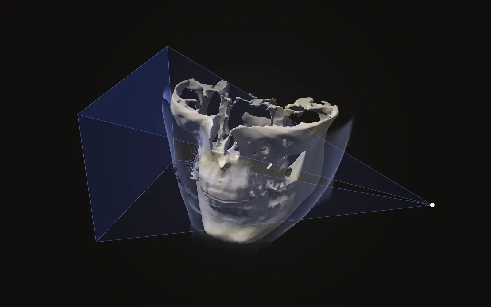
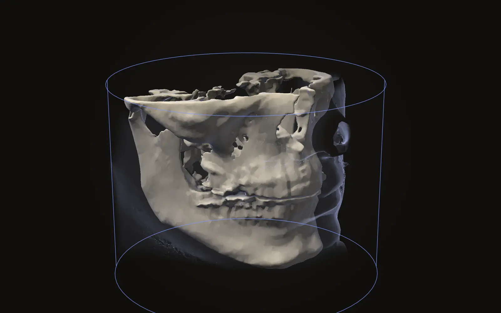
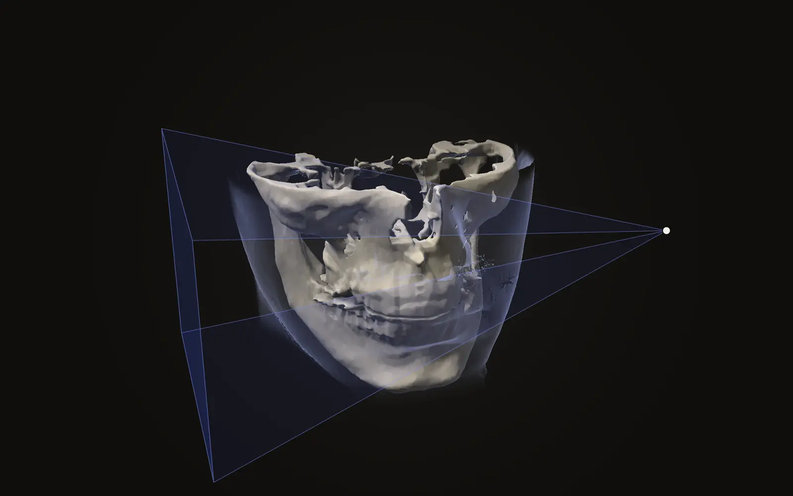
The field of view is the region the scan covers. In the 14-device study, the authors concluded that small-, medium- and large-field scanners should be separated, because dose was strongly related to field size.[1] What a field of view does and does not show is covered in CBCT field of view explained.
In the 2015 meta-analysis, adult dose ranges for small, medium and large fields overlapped heavily.[2] The overlap is the reason a field label on its own tells you little about the dose of a particular scan.
Lever 2: where the field sits#
Two scans with the same field size can still differ with the height of the field on the head. A study in a 10-year-old phantom used one scanner at its large field of view. Moving the field from a cranial to a caudal position, without shielding, raised effective dose by 18.8 percent. Adding shielding in the caudal position cut the dose by 31.6 percent.[3]
The thyroid's local absorbed dose had the largest effect on the calculated effective dose, followed by the oesophagus, bone marrow and bone surfaces.[3] That fits the organ shares measured across 14 devices, where the thyroid took 21 percent.[1]
Lever 3: exposure settings, voxel size and trajectory#
A systematic review of 16 implant-dentistry studies asked how low-dose protocols are made. The studies reported five routes: lower tube voltage (kV), lower tube current (mA), lower resolution through a larger voxel size, shorter exposure time, and a different scanning trajectory.[4]
| Lever | How a low-dose protocol uses it | Source |
|---|---|---|
| Tube voltage (kV) | Reduced in some protocols; the one parameter the reviewers left out when calling reductions feasible | [4] |
| Tube current (mA) | Reduced | [4] |
| Voxel size | Larger voxels, so lower resolution | [4] |
| Exposure time | Shorter | [4] |
| Scan trajectory | Altered | [4] |
The reviewers found that low-dose protocols performed similarly to higher-resolution ones for objective measurements, with the adverse effect mostly on subjective ratings of image quality.[4] They also say clinical studies are still needed before the results can be extended to patient care, and every included study was about implant dentistry.[4] How voxel size affects sharpness is in CBCT voxel size and sharpness.
Why there is no single dose number#
A 2012 phantom study of CBCT found the dose distribution can be asymmetrical, because of an off-centre isocentre or a partial rotation of the X-ray source. It also found that dose outside the primary beam drops by about 90 percent.[5] The authors concluded that no optimal dose index is available for CBCT, because of its complicated exposure geometry and the practical side of quality-control measurement.[5]
That is one reason CBCT doses are quoted from phantom measurements and per-protocol calculations rather than read off a standard figure. For how CBCT and medical CT differ in how they are built, see CBCT versus medical CT. For a panoramic X-ray, which works differently again, see CBCT versus a panoramic X-ray.
What these studies do not tell you#
Most of the figures above come from phantoms, which stand in for a head and neck. They show what a protocol deposits in organs under test conditions. They do not show the dose of any given scan on a given day. The two 2012 studies and the 2022 study measured specific machines, and the 2024 review covered only implant dentistry. Machines and protocols have kept changing since the earliest of them.
Why do two CBCT machines give different radiation doses?
Machines differ in exposure factors, the diameter and height of the beam, and where the beam sits relative to radiosensitive organs. Fourteen devices measured in one phantom study ranged from 19 to 368 microsieverts.
Does a smaller field of view always mean less dose?
Usually, but not always. In a 2015 meta-analysis the adult ranges overlapped between small, medium and large fields, so a setting on one machine can deliver more than another machine's larger field.
Does voxel size change CBCT radiation dose?
A systematic review of low-dose implant protocols lists larger voxel size, meaning lower resolution, among the ways doses were reduced, alongside tube voltage, tube current, exposure time and scan trajectory.
Is there a single number that describes CBCT dose?
Not yet. A 2012 phantom study concluded that no optimal dose index exists, because the exposure geometry is complicated and quality-control measurement is hard to do in practice.
Sources
- Pauwels R, Beinsberger J, Collaert B, et al. (2012). Effective dose range for dental cone beam computed tomography scanners. Eur J Radiol, 81: 267-71. Two anthropomorphic phantoms with thermoluminescent detectors, 14 devices, ICRP 2007 weights.DOIPubMed
- Ludlow JB, Timothy R, Walker C, et al. (2015). Effective dose of dental CBCT, a meta analysis of published data and additional data for nine CBCT units. Dentomaxillofac Radiol, 44: 20140197. Meta-analysis of 20 publications plus new measurements on nine units.DOIPubMed
- Vogiatzi T, Menz R, Verna C, et al. (2022). Effect of field of view (FOV) positioning and shielding on radiation dose in paediatric CBCT. Dentomaxillofac Radiol, 51: 20210316. One scanner, large field of view, a 10-year-old phantom.DOIPubMed
- Kaaber L, Matzen LH, Schropp L, et al. (2024). Low-dose CBCT protocols in implant dentistry: a systematic review. Oral Surg Oral Med Oral Pathol Oral Radiol, 138: 427-39. Sixteen studies, all in implant dentistry.DOIPubMed
- Pauwels R, Theodorakou C, Walker A, et al. (2012). Dose distribution for dental cone beam CT and its implication for defining a dose index. Dentomaxillofac Radiol, 41: 583-93. Water and PMMA head-size phantoms.DOIPubMed
Terms used
- Effective dose
- A single number in sieverts that adds up the dose to each organ, weighted by how sensitive that organ is to radiation. It lets you compare different examinations.
- Field of view
- The size of the region a scan covers. A small field of view shows a few teeth; a large one shows the whole head.
- Voxel
- A three-dimensional pixel. A scan is a grid of voxels, each with one grey value.
This is general information, not medical advice. Jaw Viewer is not a medical device. Talk to your surgeon, orthodontist or radiologist about your own case.