Key points
- The field of view is the region a CBCT scan captures. Anything outside it was never recorded, so no software can show it later.
- In a 2015 meta-analysis, mean adult effective dose was 84 microsieverts for small fields of view, 177 for medium and 212 for large.
- Doses for the same field size vary widely between machines: reported adult ranges overlap, from 5 to 652 microsieverts for small fields and 46 to 1073 for large.
- Reviews of CBCT scans found at least one incidental finding in 24.6 to 94.3 percent of scans, and most lay outside the tooth-bearing region.
A CBCT scan covers only the region the scanner was set to capture, called the field of view. A small one can hold a few teeth and the bone around them, and a large one can take in most of the head. Whatever sits outside that region was never recorded, so it is not in the files.
What does field of view mean?#
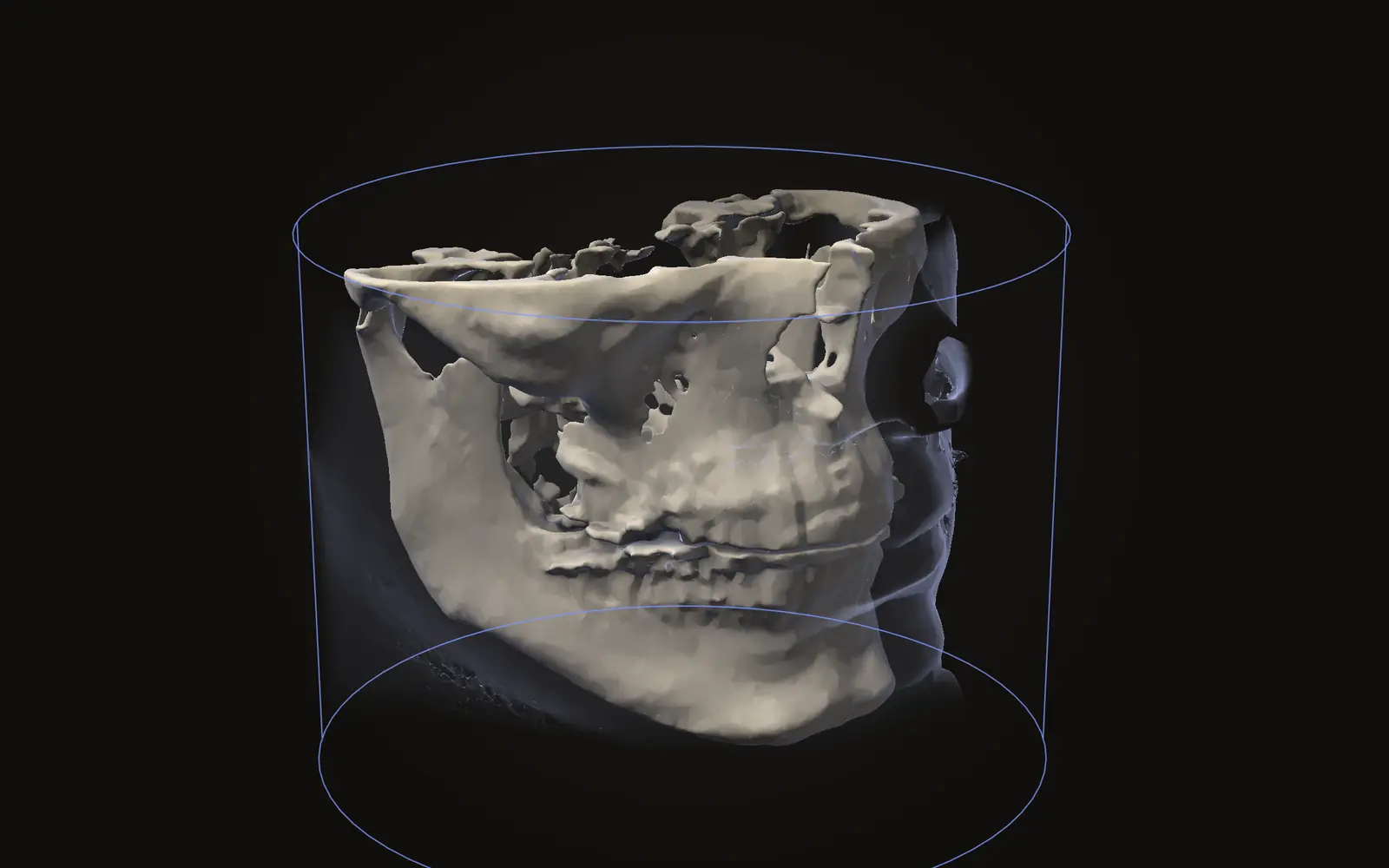
Field of view (FOV) is the size of the region a scan covers. On a CBCT scanner, the geometric settings of the acquisition influence both image quality and patient dose.[1] The field of view is one of them, and the result is a block of data. Inside it, every voxel has a value. Outside it, there is nothing.
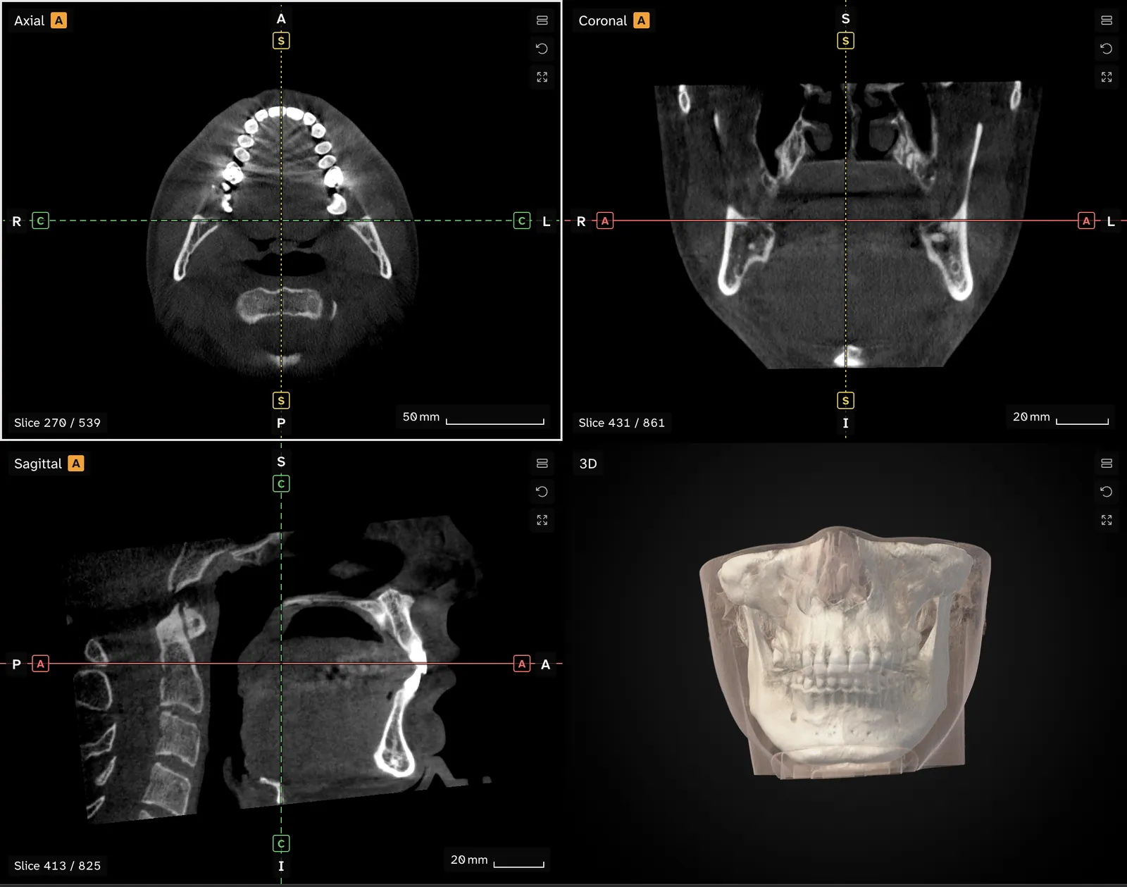
This is why two scans of two people can look so different when you open them. One may show a single quadrant of teeth. Another may show the whole lower face, the neck bones and the base of the skull. Both are normal CBCT scans, taken to answer different questions.
Why is the scan cut off?#
When researchers pooled published dose measurements, mean adult effective doses grouped by field size were 84 microsieverts for small fields, 177 for medium and 212 for large.[2] For children the pooled means were 103 microsieverts for small fields and 175 for large and medium fields combined.[2]
On average, then, a narrower field goes with a lower dose, and it keeps the picture on one region. The operator chooses the field before the scan. Which choice suits which question is for the clinician, and this article does not try to judge it.
How much does the dose vary?#
Field size is only one factor affecting dose. The ranges overlap heavily.
| Field of view | Adult range, any protocol | Mean, standard protocols |
|---|---|---|
| Small | 5 to 652 µSv | 84 µSv |
| Medium | 9 to 560 µSv | 177 µSv |
| Large | 46 to 1073 µSv | 212 µSv |
Source: Ludlow et al., 2015.[2]
The same review reported large differences between CBCT units, and noted that low-dose and high-definition protocols available on many units extend the range further.[2] A small field on one machine can deliver more than a large field on another. The full breakdown of what drives dose is in why one CBCT scan delivers more than another, and the comparison with medical CT is in CBCT versus CT radiation dose.
What is missing from a small field of view?#
Missing anatomy limits what you can do in a viewer:
- Airway volume and sinus volumes can only be measured if the whole airway or sinus is inside the scan.
- A skull-base landmark, or a distance that runs to the edge of the head, needs the region to be there.
- Slices in all three directions end at the same boundary. In the axial, coronal and sagittal views you will see the image stop at the edge of the captured block.
Does field of view change the numbers on the image?#
It can. The grey values of a CBCT scan are not calibrated, and one review lists the limited field size, scattered radiation and limits of the reconstruction algorithms among the reasons grey values vary from scan to scan.[3] The same authors conclude that quantitative use of grey values in CBCT should generally be avoided at this time.[3] For that reason, comparing a grey value from a small-field scan with one from a large-field scan is not a like-for-like comparison. More on this in what a CBCT scan shows and cannot tell you.
What about things outside the area of interest?#
A large field captures more than one region, and reviewers have looked at what else turns up. A 2013 systematic review of five studies found that between 24.6 and 93.4 percent of CBCT scans contained incidental findings, and that the majority were outside the region of the teeth and their supporting bone.[4] A 2019 review of ten studies reported a similar range, 24.6 to 94.3 percent, and linked incidental findings with the frequent use of a large field of view.[5] It also reported that threatening findings were rare, at 1.4 percent.[5]
Those percentages describe groups of scans, and they vary a lot between studies. They say nothing about any one person's scan. What the incidental findings are, and how often they matter, is covered in incidental findings on CBCT scans.
How to see the extent of your own scan#
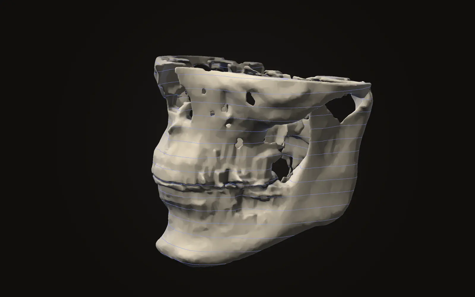
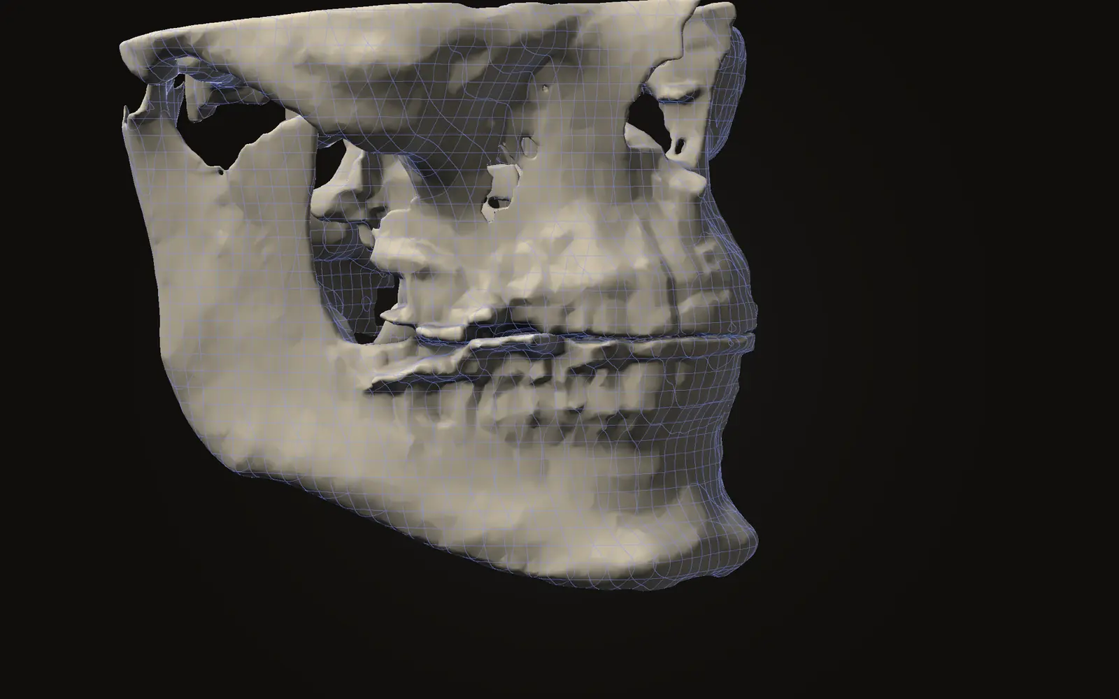
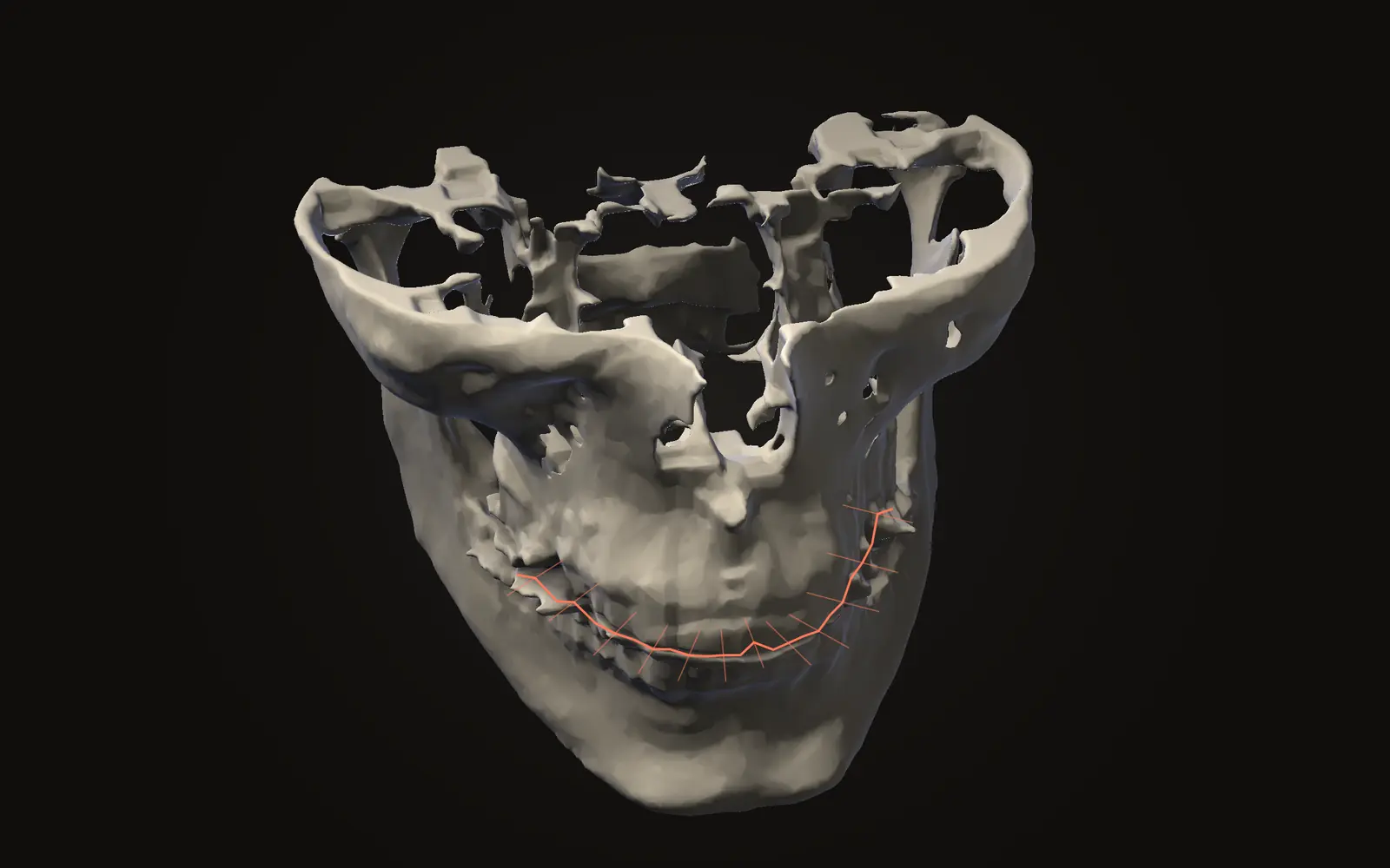
Open the scan in Jaw Viewer and the 3D view shows the shape of the captured data. Turn it and you can see where it begins and ends. Scroll the slices and the picture stops at the same boundary. The step-by-step is in the guide on opening your scan, and the tools for looking around are in view and measure.
The viewer shows what is in the files. It cannot reconstruct anatomy that was never captured, and it does not guess what lies beyond the edge.
Common questions#
Why does my CBCT scan show only my teeth and jaw?
The scanner was set to capture a limited region, called the field of view. Only that region is recorded in the files. Anything outside it was never imaged, so it cannot be added back afterwards.
Does a bigger field of view mean more radiation?
On average, yes. A 2015 meta-analysis put mean adult effective dose at 84 microsieverts for small fields, 177 for medium and 212 for large. The machine and protocol change the dose a lot as well.
Can the viewer show parts of the head that are not in the scan?
No. The viewer only displays what is in the files you open. If the airway, sinuses or skull base lie outside the field of view, they are not there to show or measure.
Sources
- Scarfe WC, Farman AG (2008). What is cone-beam CT and how does it work?. Dent Clin North Am, 52: 707-30.DOIPubMed
- Ludlow JB, Timothy R, Walker C, et al. (2015). Effective dose of dental CBCT, a meta analysis of published data and additional data for nine CBCT units. Dentomaxillofac Radiol, 44: 20140197. 20 publications after screening, plus data for nine units.DOIPubMed
- Pauwels R, Jacobs R, Singer SR, et al. (2015). CBCT-based bone quality assessment: are Hounsfield units applicable?. Dentomaxillofac Radiol, 44: 20140238.DOIPubMed
- Edwards R, Altalibi M, Flores-Mir C (2013). The frequency and nature of incidental findings in cone-beam computed tomographic scans of the head and neck region: a systematic review. J Am Dent Assoc, 144: 161-70. Five studies met the inclusion criteria.DOIPubMed
- Dief S, Veitz-Keenan A, Amintavakoli N, et al. (2019). A systematic review on incidental findings in cone beam computed tomography (CBCT) scans. Dentomaxillofac Radiol, 48: 20180396. Ten studies, 90 to 1000 participants.DOIPubMed
Terms used
- Field of view
- The size of the region a scan covers. A small field of view shows a few teeth; a large one shows the whole head.
- CBCT
- Cone beam computed tomography. A scanner that rotates once around the head and records a cone-shaped X-ray beam, common in dentistry and jaw surgery.
- Voxel
- A three-dimensional pixel. A scan is a grid of voxels, each with one grey value.
This is general information, not medical advice. Jaw Viewer is not a medical device. Talk to your surgeon, orthodontist or radiologist about your own case.