Key points
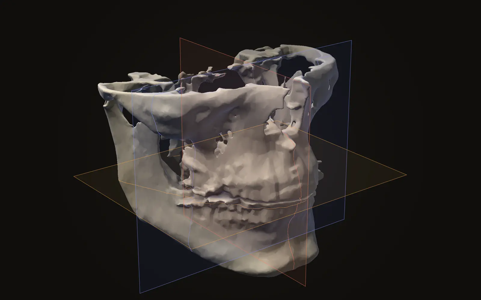
- Axial, coronal and sagittal are three slice directions through the same scan: axial cuts across the head, coronal splits front from back, sagittal splits left from right.
- A scan is one 3D block, so the three views are not three scans. Moving the crosshair in one view moves the matching point in the other two.
- DICOM patient axes: x increases toward the patient's left, y toward the back, z toward the head. Each slice file stores its position and orientation in these axes.
- Letters in the corners of a slice name the patient's directions. Read them instead of assuming which side of the screen is the left side of the patient.
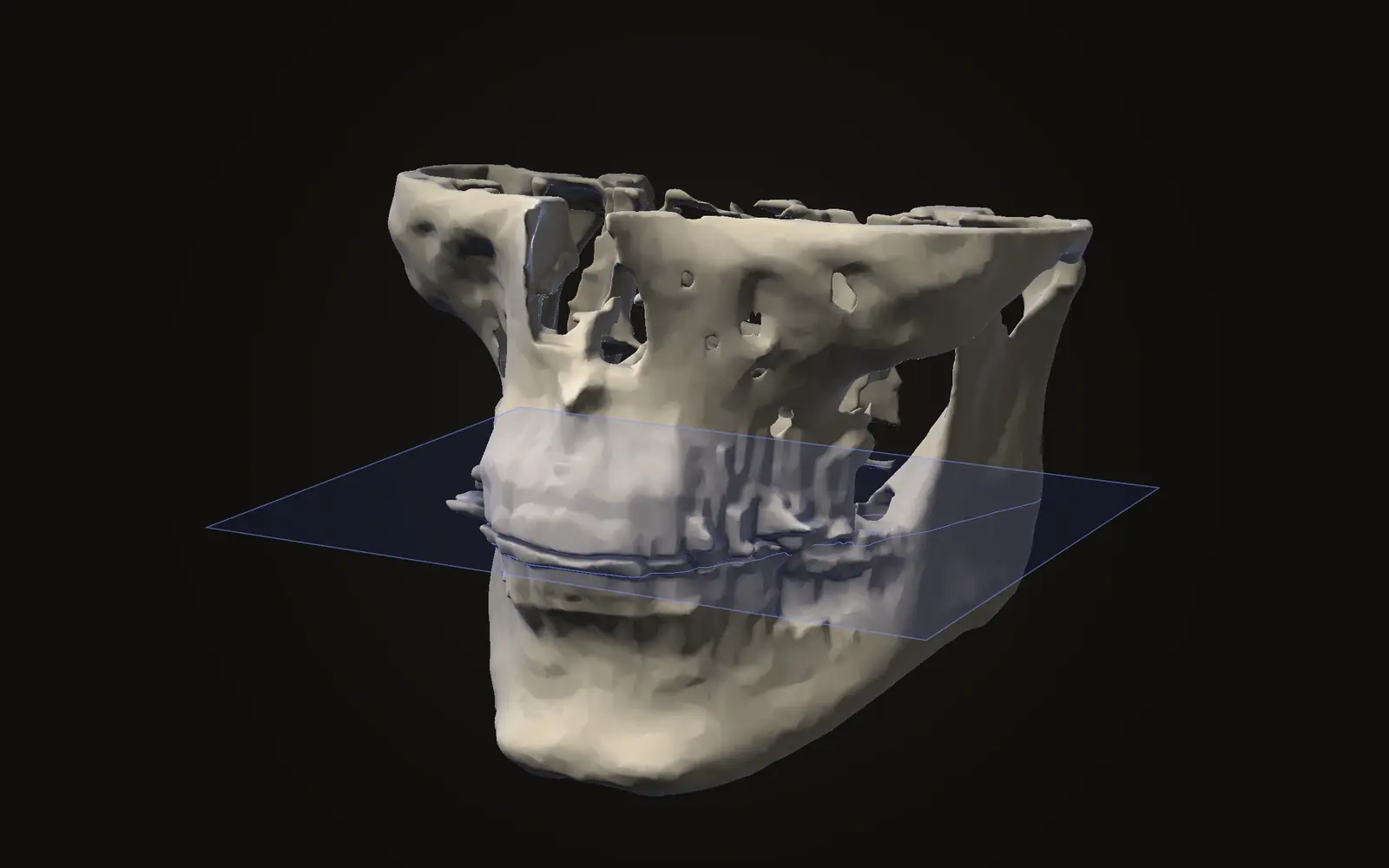
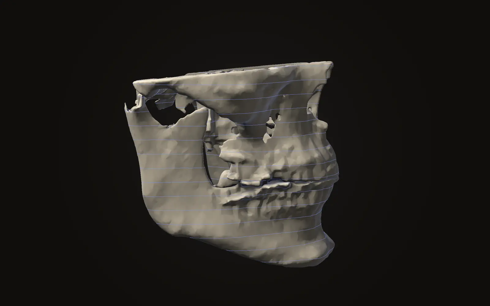
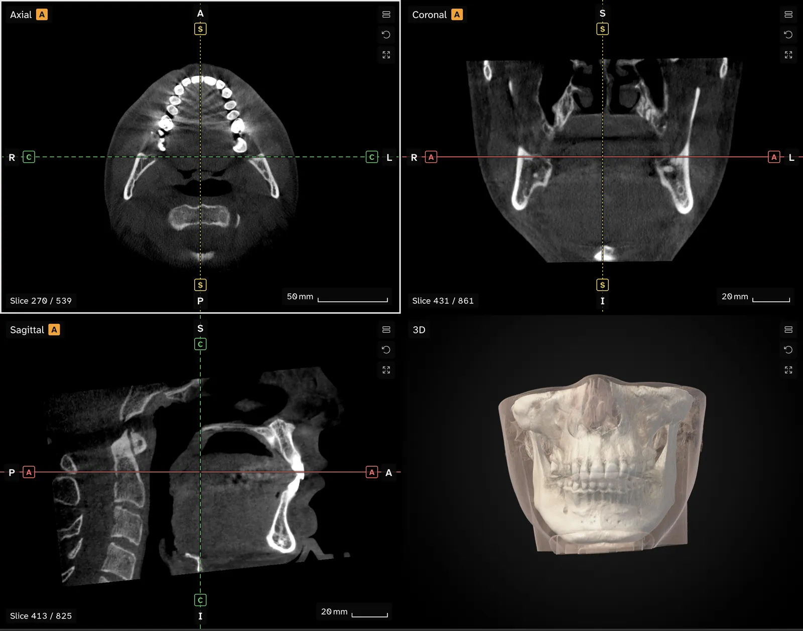
Axial, coronal and sagittal are the three standard directions in which a scan is sliced. Axial slices cut across the head, coronal slices separate the front from the back, and sagittal slices separate the left from the right. All three come from the same scan, so the same spot appears in each of them.
What are the three directions?#
Picture the head standing upright. An axial slice is a horizontal cut, as if you looked down on a disc of the head from above. A coronal slice runs from ear to ear, so you look at the face from the front with the front part of the head peeled away. A sagittal slice runs from front to back, so you see the head in profile, with half of it removed.
DICOM, the standard that scan files follow, draws all three planes in its guide to patient orientation, under the names transverse, sagittal and coronal.[1, Figures A-5 to A-7] Scan viewers, including Jaw Viewer, use the word axial for the transverse plane.
| View | Cuts across | You see |
|---|---|---|
| Axial | Top to bottom | A horizontal disc, from above |
| Coronal | Front to back | The face from the front |
| Sagittal | Left to right | The head in profile |
Why one scan gives three views#
A CBCT scanner records one block of voxels, not three separate sets of pictures. The files are stored as a stack of slices, and each file carries its own position and orientation in the patient. The DICOM Image Plane module holds the position of the slice corner, the directions of its rows and columns, and the spacing between pixel centres in millimetres.[2, C.7.6.2] Software uses these details to place every slice in a shared 3D space and cut through it in any direction.
So the other views are computed from the same stack of slices. Nothing is added: each coronal or sagittal image is assembled from voxels the scan already holds, and it can only be as sharp as those voxels are.
Which way is left?#
DICOM fixes the patient's axes. The x axis increases toward the patient's left, the y axis toward the patient's back, and the z axis toward the head.[2, C.7.6.2] Those axes belong to the patient, not to the screen.
Viewers differ in how they draw slices on screen. Read the letters in each corner for the patient's directions, as the Jaw Viewer viewing guide explains. If you switch from one viewer to another and a slice looks mirrored, check those letters before drawing a conclusion about the scan.
How do the three views work together?#
Because the views share one space, they stay linked. In Jaw Viewer you focus a slice view and scroll to move through it, click a point to place the crosshair, and find that point in the other two views. A feature that is faint in one direction is often clear in another, which is why a measurement is worth checking in more than one view.
A few habits make the three views easier to read:
- Start with the axial view for the shape of the dental arch, then click a tooth or a canal and look at it from the side in the sagittal view.
- Use the coronal view to compare the left and right sides at the same height.
- Scroll slowly, and check a point in all three views before you trust it.
The viewer also draws a 3D surface from the scan. Check a point in the slices before you rely on a measurement placed on that surface.
Slices that are not axial, coronal or sagittal#
Teeth, the jaw and the nerve canal do not line up with the three standard directions. An oblique slice is cut at a tilt, so it can run along the length of one tooth or across the jaw at a right angle to its curve. Jaw Viewer offers slices in three directions and oblique slices.
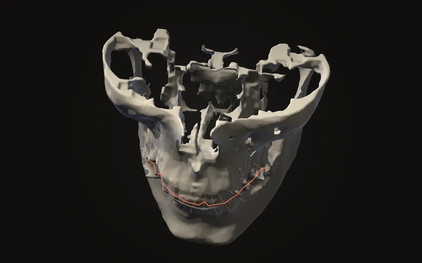
A curved cut follows the dental arch and is flattened into a panoramic image. It is a computed view of the scan you already have, as the article on the panoramic image from a CBCT explains. A panoramic X-ray taken in a separate exposure is a different examination; the comparison is in CBCT versus panoramic X-ray. The panoramic views guide covers how to set up the curve.
Where to go from here#
If you are still getting oriented, what a CBCT scan shows describes the structures you will meet in each view. If a slice looks too bright or too grey, the article on window and level explains the display setting. For how the slices are stored and where the position data sits in each file, see what is inside a DICOM file.
Are axial, coronal and sagittal three different scans?
No. They are three ways of slicing one scan. The scanner records a single 3D block, and the software cuts it along each of the three directions.
What does the letter in the corner of a slice mean?
It names a direction in the patient, such as R for right, L for left, A for anterior (front) or P for posterior (back). It tells you which way you are looking.
Is axial the same as transverse?
Both words describe a slice across the body at right angles to the head-to-foot line. DICOM figures label it transverse, and scan viewers usually call it axial.
Can I cut a slice at a tilt?
Yes. An oblique slice is cut at any angle, for example along the length of a tooth or a jaw. The viewer offers oblique slices beside the three standard ones.
Sources
- DICOM Standards Committee (2026). DICOM PS3.17, Explanatory Information, Annex A (Explanation of Patient Orientation). NEMA. Version 2026d, read 21 November 2026. The annex shows the transverse, sagittal and coronal planes in its figures A-5 to A-7.Link
- DICOM Standards Committee (2026). DICOM PS3.3, Information Object Definitions, section C.7.6.2 (Image Plane Module). NEMA. Version 2026d, read 21 November 2026.Link
Terms used
- Axial
- A slice that cuts across the body at right angles to the head-to-foot line, like a horizontal slice through the head. Also called transverse.
- Coronal
- A slice that runs from ear to ear and splits the head into a front part and a back part.
- Sagittal
- A slice that runs from front to back and splits the head into a left part and a right part.
- CBCT
- Cone beam computed tomography. A scanner that rotates once around the head and records a cone-shaped X-ray beam, common in dentistry and jaw surgery.
- Voxel
- A three-dimensional pixel. A scan is a grid of voxels, each with one grey value.
This is general information, not medical advice. Jaw Viewer is not a medical device. Talk to your surgeon, orthodontist or radiologist about your own case.