Key points
- A panoramic image made from a CBCT scan is computed from the existing 3D data along a curve you place. It is a reformatted view, not a separate X-ray exposure.
- In 28 CBCT datasets, crown-width measurements on reformatted panoramic images differed significantly from axial measurements, and the error grew with distance from the centre of the curve.
- Against a caliper, CBCT panoramic reconstructions read 1.7 mm (SD 1.2 mm) short on tooth length in one study; conventional panoramic radiographs read 6.3 mm (SD 2.0 mm) long.
- Where the curve sits changes what the panoramic image shows, so check the curve on an axial slice and confirm a structure in the cross-sections.
A panoramic image made from a CBCT scan is a curved slice computed from the scan's 3D data, laid out flat so it resembles a panoramic X-ray. No second exposure is involved: the panoramic images in the 2026 study below were reformatted from CBCT datasets that already existed.[1] The curve’s position affects the image and the measurements you take from it. Studies have measured those errors.
How is a panoramic image made from a volume?#
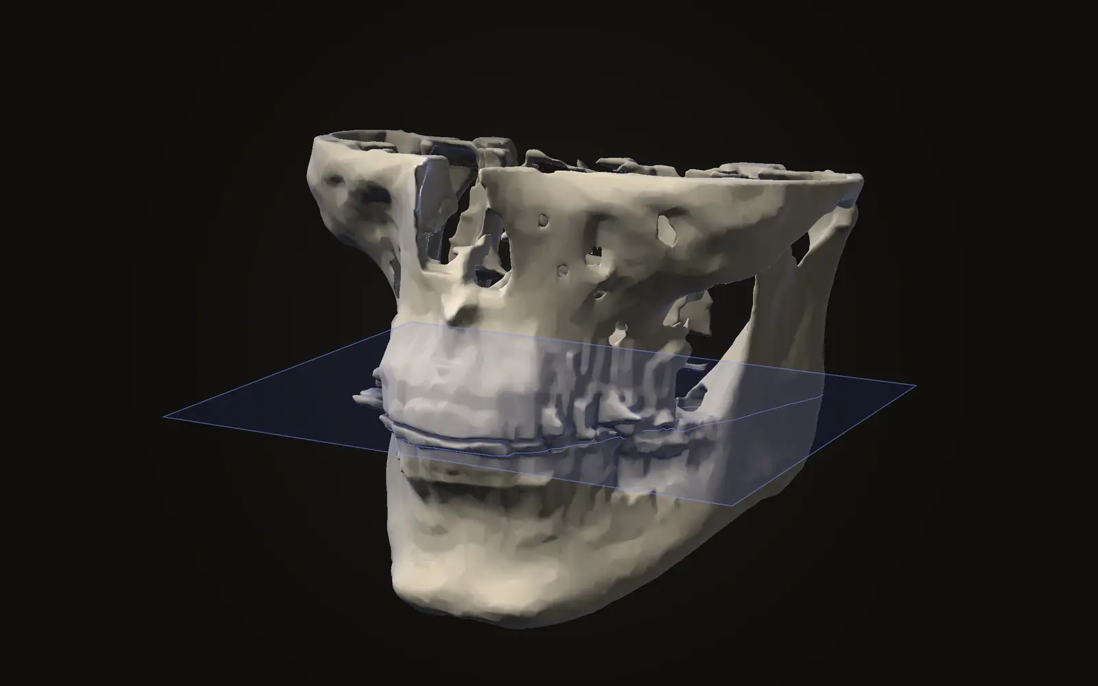
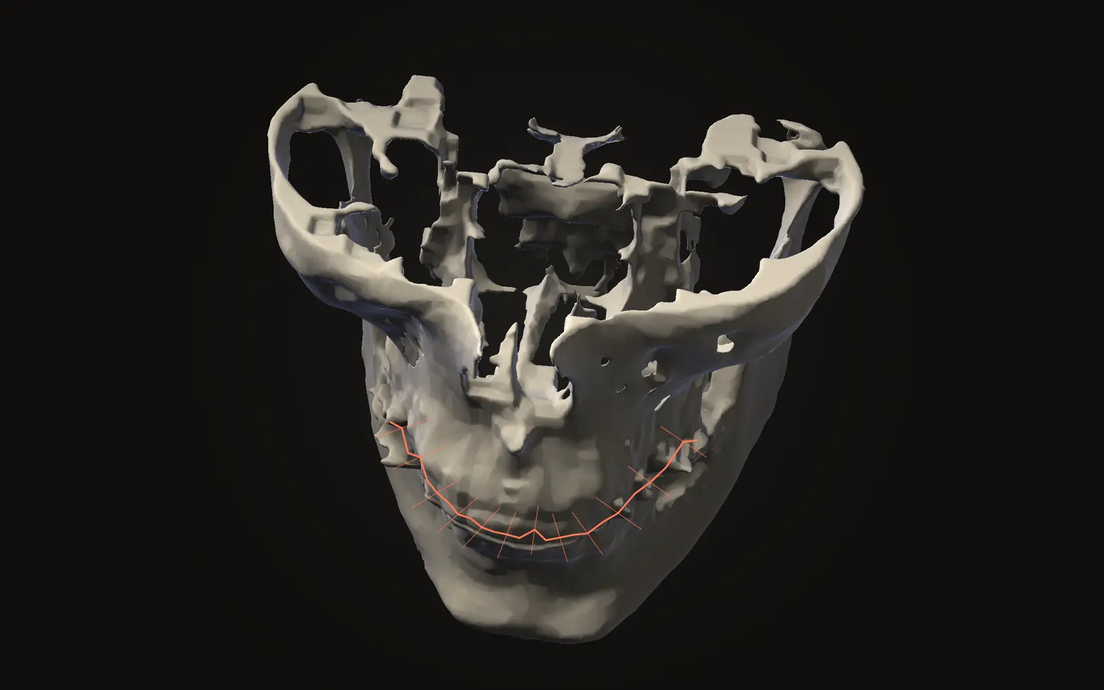
A CBCT scan is a grid of voxels. To get a panoramic-style picture, software follows a curve along the dental arch on the axial slice, samples the voxels along that path and spreads them into a flat image. The result is a panoramic reconstruction.
Software can draw the curve by itself or you can place it by hand. One 2026 study compared three ways of doing this on 28 CBCT datasets: an automatic reconstruction, a manual one oriented to the nasal spine plane, and a manual one oriented to the occlusal plane.[1] The same scan therefore gives different panoramic images depending on the workflow, and the authors reported that image quality scores differed across the three.[1]
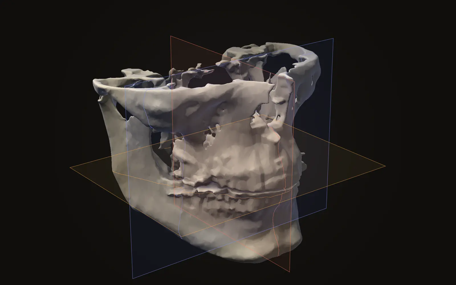
This is a different object from the flat image a dedicated panoramic machine records in one pass. That comparison, including the dose side, is in CBCT vs panoramic X-ray. The three slice directions the curve is drawn on are explained in axial, coronal and sagittal views.
How accurate are measurements on it?#
The four studies used different designs and reference methods, so their results need to be read separately.
| Study | Material | Reference | What was reported |
|---|---|---|---|
| Almeida 2021 | 10 dry mandibles, 3 software packages | Digital caliper | No statistically significant difference; relative error 1.3 to 8.0%, depending on software and voxel size[2] |
| Flores-Mir 2014 | 48 extracted teeth, 26 subjects | Digital caliper | CBCT panoramic reconstructions read tooth length 1.7 mm (SD 1.2 mm) short, about 4%; conventional panoramic radiographs read 6.3 mm (SD 2.0 mm) long, about 29%[3] |
| Lupi 2018 | 160 mandibular third molars | 3D measurement of the same scan | Angle between second and third molar differed by a mean of -2.3° ± 6.3°; absolute error 3.6° ± 5.7°, relative error 10%[4] |
| Fahd 2026 | 28 CBCT datasets | Axial slices of the same scan | Crown-level measurements differed significantly in all three workflows[1] |
The dry-mandible study found the measurements reliable, while the 2026 study did not, and both are fair readings of what they tested. Almeida and colleagues measured dry mandibles and compared against a caliper.[2] Fahd and colleagues measured crowns and the gaps between roots, and compared against axial images of the same scan.[1] A reformatted panorama can agree with a caliper on average and still be off at a particular spot.
Flores-Mir and colleagues also put the CBCT version beside an ordinary panoramic X-ray in living patients. The CBCT-derived image was the closer of the two to the caliper.[3] That is a statement about tooth length in 26 subjects, not about every measurement.
Why does the curve matter so much?#
The 2026 study defined a distance for each measurement site: how far it lay from the centre of the drawn curve. In every workflow, measurement error rose as the site moved away from that centre.[1] Sites near the centre of the curve were measured with less error than sites far from it.
The angle result is a useful example. Tilt between two molars looked different on the flat panoramic image than in 3D, by a mean of 2.3°, with a standard deviation of 6.3° across the 160 teeth.[4] If you want a number for a tooth or a bone, the slices in three directions are the place to take it. See how to measure on a CT scan and how accurate CBCT measurements are.
What does the viewer do?#
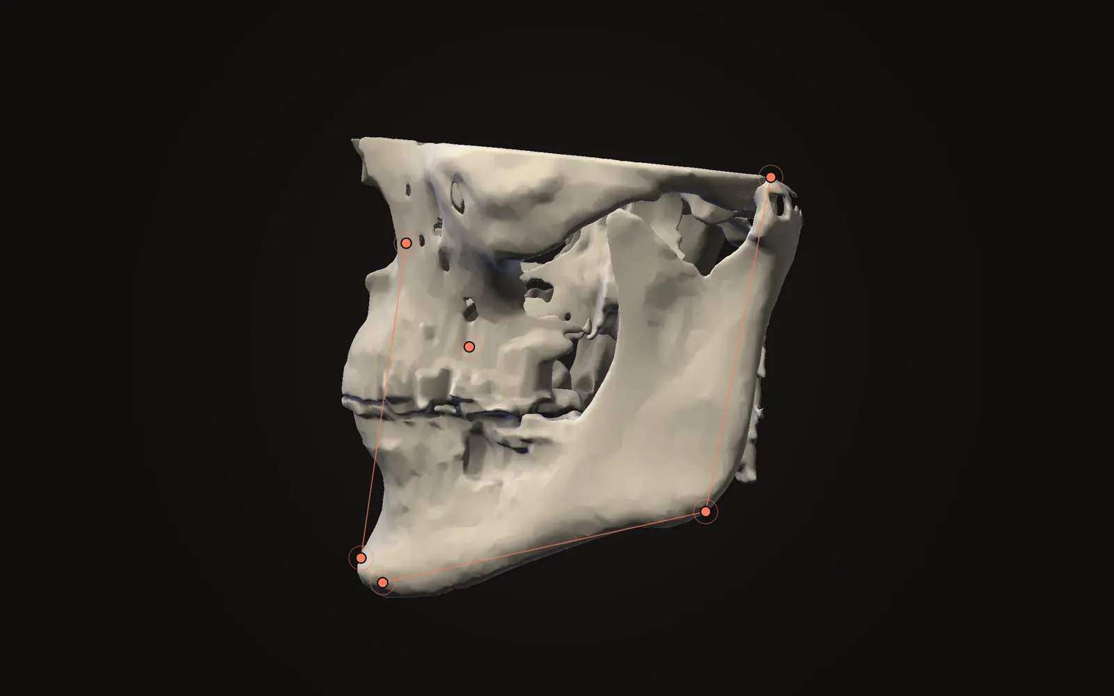
Jaw Viewer has a panoramic layout in Measure, where the nerve canal tool traces on it. You set the arch curve on the axial slice, check that it follows the teeth on both sides, and read the panoramic view together with a strip of cross-sections cut across the arch. Moving along the strip shows neighbouring cuts, and the matching spot can be found on the axial view. The panoramic views guide lists the steps.
Use the cross-section strip to confirm a structure. If a structure looks stretched or disappears on the panoramic image, the guide's first advice is to check the curve and the slice position. The nerve canal tool follows the same habit: mark the path, then check it in the cross-sections instead of trusting a smooth tube. The background to that is in the inferior alveolar nerve canal on CBCT.
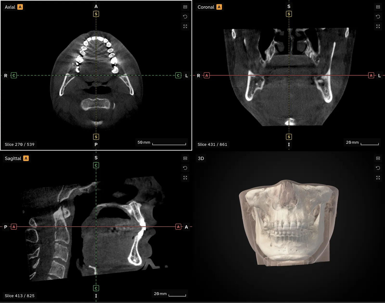
A panoramic view in the viewer is a way of looking, with the resolution and limits of the scan it comes from. Numbers from it fall under the same caution as in the studies above, and the limits page says what a measurement on screen can and cannot tell you.
Is a panoramic view from a CBCT a new X-ray?
No. It is computed from the 3D data the CBCT scan already holds, along a curve through the dental arch. No second exposure is made, and no detail is added that the scan did not record.
Why does my CBCT panoramic image look different from my panoramic X-ray?
The two are made differently. A panoramic X-ray is one exposure on a moving machine. A CBCT panoramic image is a curved slice through a volume, so it depends on where the curve is placed.
Can I measure teeth on a CBCT panoramic image?
Studies report errors that depend on the software, the voxel size and how far the site lies from the curve. One 2026 study advised cross-checking against other views before relying on a number.
What does the arch curve do?
It sets the path of the slice. Anatomy near the curve is shown most faithfully, and a misplaced curve can stretch, shrink or hide structures.
Sources
- Fahd A, Ellabban MT, Bornstein MM, et al. (2026). A comparison of panoramic views reformatted from CBCT scans using manual or automated workflows: an evaluation of geometric accuracy and subjective image quality. Clin Oral Investig, 30: 414. 28 CBCT datasets, three reconstruction workflows.DOIPubMed
- Almeida VSM, Bomfim RT, Sobreira ACR, et al. (2021). Linear measurement accuracy of CBCT panoramic reconstructions: experimental study with dry human mandibles. Oral Radiol, 37: 421-426. Ten dry mandibles, three software packages, caliper as reference.DOIPubMed
- Flores-Mir C, Rosenblatt MR, Major PW, et al. (2014). Measurement accuracy and reliability of tooth length on conventional and CBCT reconstructed panoramic radiographs. Dental Press J Orthod, 19: 45-53. 48 extracted teeth from 26 subjects, digital caliper as reference.DOIPubMed
- Lupi SM, Galinetto P, Cislaghi M, et al. (2018). Geometric distortion of panoramic reconstruction in third molar tilting assessments: a comprehensive evaluation. Dentomaxillofac Radiol, 47: 20170467. 160 mandibular third molars, 2D panoramic reconstruction against 3D measurement.DOIPubMed
Terms used
- CBCT
- Cone beam computed tomography. A scanner that rotates once around the head and records a cone-shaped X-ray beam, common in dentistry and jaw surgery.
- Voxel
- A three-dimensional pixel. A scan is a grid of voxels, each with one grey value.
- Axial
- A slice that cuts across the body at right angles to the head-to-foot line, like a horizontal slice through the head. Also called transverse.
- Panoramic reconstruction
- A curved slice that follows the dental arch, computed from an existing CBCT scan. It resembles a panoramic X-ray without a second exposure.
This is general information, not medical advice. Jaw Viewer is not a medical device. Talk to your surgeon, orthodontist or radiologist about your own case.