Key points
- A scan stores 12 to 16 bits per pixel, which is 4,096 to 65,536 grey shades, while many medical screens showed only 256. A window chooses which range of values fills the screen.
- Window level is the middle of the range and sets overall brightness. Window width is the size of the range and sets contrast. A narrow window gives strong contrast, a wide one looks flat.
- In a chest CT phantom study, three standard windows gave different sizes for the same 5.8 to 21.4 mm test nodules, so the edge you see depends on the window.
- CBCT grey values cannot be converted to Hounsfield units according to one systematic review, so window numbers from a medical CT do not carry over to a CBCT scan.
A scan looks too bright or too grey because the screen can only show part of the grey range the scan stores, and the window decides which part. Window level moves that range up or down, which changes brightness. Window width stretches or squeezes it, which changes contrast.
Why does a scan need a window at all?#
Scanners record far more shades of grey than a screen displays. Medical images typically hold 12 to 16 bits per pixel, which is 4,096 to 65,536 shades, and for a long time medical LCDs supported only 8 bits, or 256 shades.[1] Showing all of that on one screen would squeeze it into too few steps, so software picks a range and spreads it from black to white.
That range is the window. Values below it are drawn black and values above it are drawn white. The DICOM standard describes this as selecting the range of input values that are mapped to the full range of the display.[2, C.11.2.1.2] Values outside the window stay in the file. Move the window to see them.
What do level and width do?#
The standard defines level as the input value at the centre of the window, which it calls Window Center, and width as the size of the range.[2, C.11.2.1.2.1] The two settings are stored with the image as a suggested display, and a file can hold more than one pair.[2, C.11.2] Read more about those header attributes in what is inside a DICOM file.
The settings explain several common display problems:
- A slice that looks pale and flat usually has a window much wider than the values that matter, so everything lands in the same few mid greys.
- A slice that looks dark or has large black areas has a level set too high, so most values fall below the window.
- A slice that looks blown out has a level set too low, so most values fall above it.
A narrow window gives strong contrast between values inside it, and it clips everything else to black or white. Reducing the width can make a thin edge look sharp or a faint structure disappear.
How do the usual windows differ?#
Textbook windows have names such as soft tissue and lung. The chest CT study below gives a concrete example. It used three standard settings in Hounsfield units, and the range each one shows follows from level plus or minus half the width.[3][2, C.11.2.1.2.1]
| Setting | Width | Level | Range shown |
|---|---|---|---|
| Soft tissue | 400 HU | +20 HU | -180 to +220 HU |
| Lung | 850 HU | -750 HU | -1,175 to -325 HU |
| Broad lung | 1,350 HU | -550 HU | -1,225 to +125 HU |
The soft tissue window shows a narrow band around water. The lung windows sit far lower, around the values of air-filled tissue, so a structure of soft tissue density would be white and lose its inner detail. Each window is a different picture of the same data.
Does the window change how big something looks?#
It can. In that study the authors scanned 45 synthetic nodules of 5.8 to 21.4 mm and measured each at the three settings. Measurements at either lung setting were highly accurate, while measurements at the soft tissue setting were inaccurate, and the authors concluded that window choice matters when CT is used to size nodules.[3]
This was a chest CT phantom, so it says nothing about jaw scans or about any particular dental measurement. It does show the general mechanism: where an edge appears depends on which values the window turns black or white. When you measure on a slice, the window is one of the things that moves the edge you are looking at.
How does this work in the viewer?#
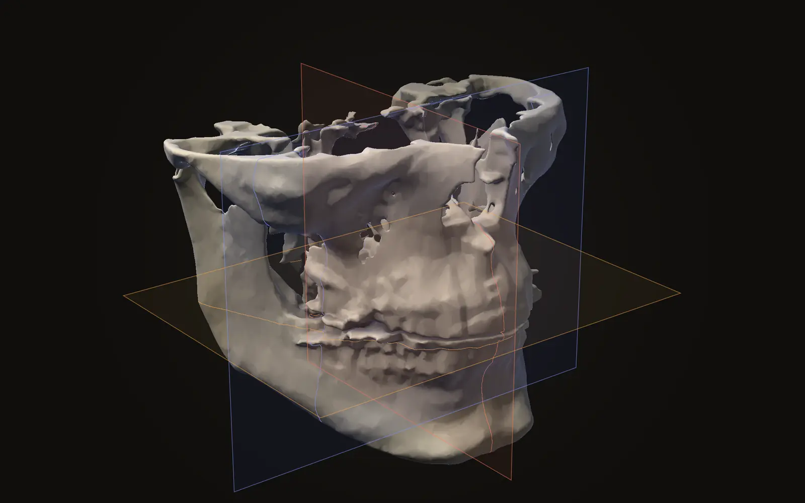
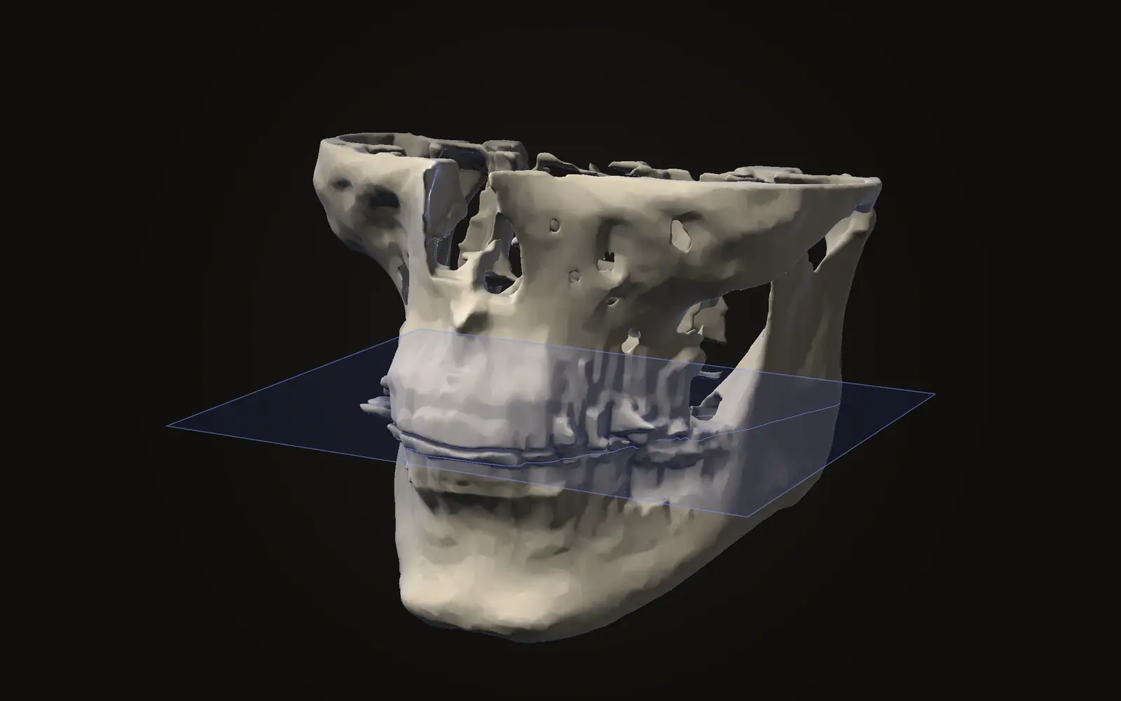
Jaw Viewer calls level Brightness and width Contrast. They apply to the slice views, which are the three directions described in axial, coronal and sagittal views explained.
- Pick the Brightness/contrast tool and drag on a slice. The setting applies to every slice view of that scan.
- The Slices section of the inspector has a preset list: From scan, Bone, Teeth, Soft tissue and Airway.
- The same section has a small pad and two number fields. Left and right on the pad change brightness, up and down change contrast, and a double-click on the pad returns to From scan.
From scan uses the display window stored in the file when there is one.[2, C.11.2] The other presets are calculated from the grey values of the scan you opened, so treat them as starting points for that scan. None of this edits the scan; the files are only read. The view and measure guide lists the tools, and controls and shortcuts has the keys.
Why does my CT or CBCT scan look washed out?
The window is probably wide or centred away from the structure you want. Narrowing the width adds contrast, and moving the level shifts brightness. The scan data itself is unchanged by either.
What is the difference between window width and window level?
Level is the grey value at the middle of the displayed range and sets overall brightness. Width is how many grey values are spread from black to white and sets contrast.
Does changing the window change my scan?
No. The window only changes how stored values are drawn on screen. The values in the files stay as they are, and in Jaw Viewer the files are never modified.
Can I use the bone window numbers from a medical CT on a CBCT scan?
Not directly. CBCT grey values are not Hounsfield units, and a systematic review found they cannot be converted. Use a preset or adjust by eye on the scan in front of you.
Sources
- Kimpe T, Tuytschaever T (2007). Increasing the number of gray shades in medical display systems, how much is enough?. J Digit Imaging, 20: 422-432. Abstract read. Written at a display manufacturer; used only for the bit depth and display figures.DOIPubMed
- DICOM Standards Committee (2026). DICOM PS3.3, Information Object Definitions, section C.11.2 (VOI LUT Module). NEMA. Version 2026d, read 21 November 2026. Window Center (0028,1050) and Window Width (0028,1051).Link
- Harris KM, Adams H, Lloyd DC, et al. (1993). The effect on apparent size of simulated pulmonary nodules of using three standard CT window settings. Clin Radiol, 47: 241-244. Abstract read. Chest CT phantom study with 45 synthetic nodules, not a dental study.DOIPubMed
- Eguren M, Holguin A, Diaz K, et al. (2022). Can gray values be converted to Hounsfield units? A systematic review. Dentomaxillofac Radiol, 51: 20210140. Abstract read. Only three studies met the inclusion criteria.DOIPubMed
Terms used
- Window level
- The grey value in the middle of the window. Moving it makes the slice look brighter or darker overall. DICOM calls it Window Center.
- Window width
- The range of grey values shown from black to white in a slice. A narrow window gives strong contrast over a small range; a wide one shows many values with less contrast.
This is general information, not medical advice. Jaw Viewer is not a medical device. Talk to your surgeon, orthodontist or radiologist about your own case.