Key points
- A DICOM file starts with a 128-byte preamble and the four letters DICM, then a header of labelled attributes, then the pixel data. One file holds one image.
- Voxel size comes from the header: Pixel Spacing gives the in-slice size in mm, and the distance between slice positions gives the third side.
- The header has a place for the patient's name and birth date, so the file can identify a person even when its file name is just I0000001.
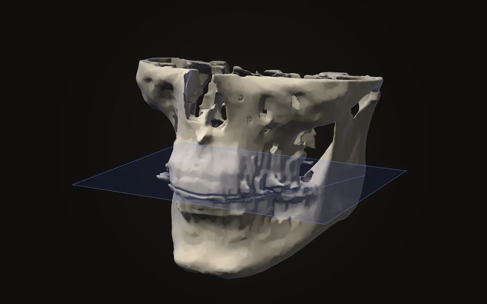
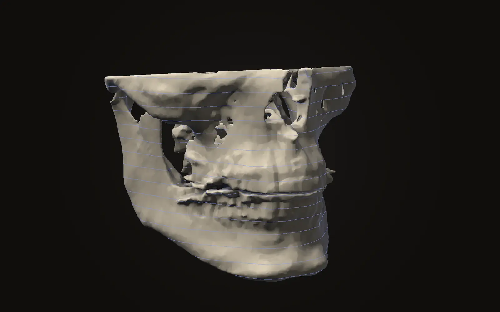
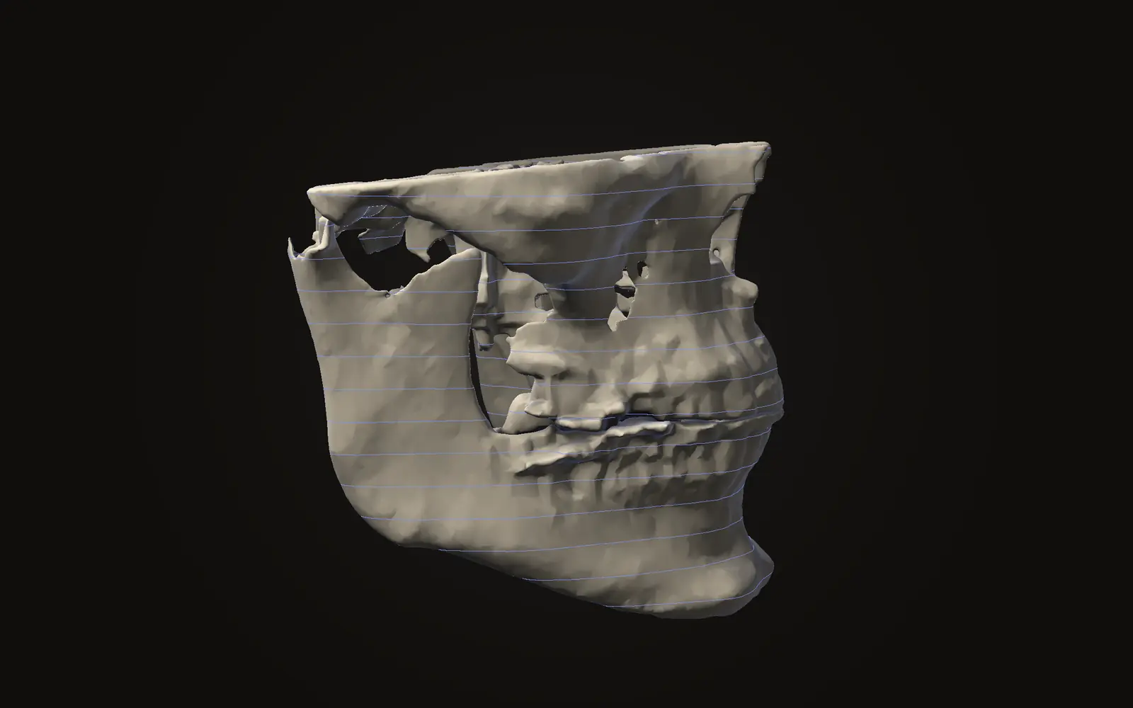
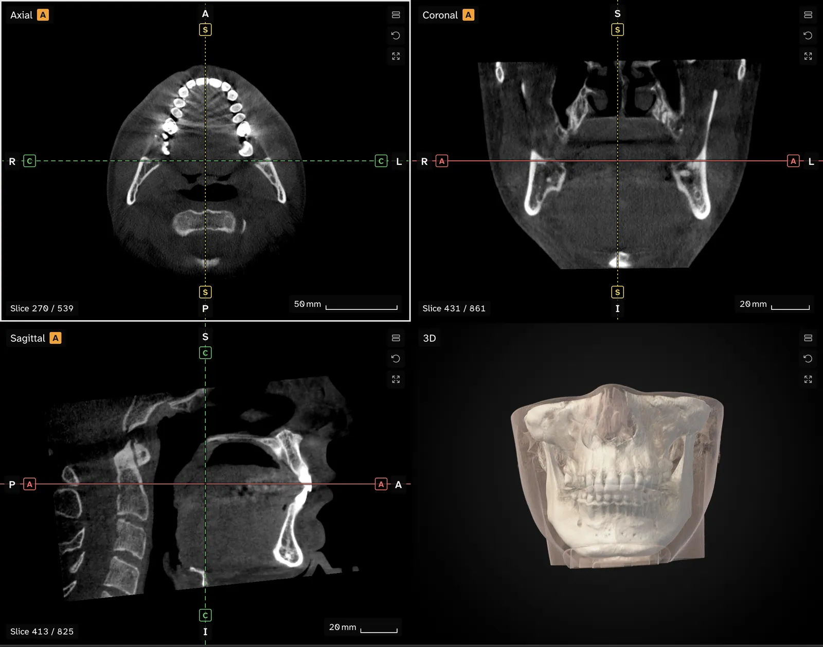
- A CBCT scan is usually hundreds of DICOM files, one per slice, that software stacks into a 3D volume using the positions stored in each header.
A DICOM file is one medical image with a header in front of it. The header is a list of labelled attributes, such as the image size, the voxel size and who the scan is of, and the pixel data follows it.[1, §7.1] A CBCT scan is hundreds of these files, one per slice.
How is the file laid out?#
DICOM was first used to pass image data between machines from different makers.[2] The header is where that information travels with the image.
Every file starts with a 128-byte preamble, which is set to zeros when nothing uses it. Then come the four letters DICM, then the header.[1, §7.1] Software uses DICM to recognise a DICOM file, and the standard tells readers not to rely on the preamble for that.[1, §7.1] The file name plays no part. This is the reason a file called I0000001 with no ending is still a valid scan slice, which the article on opening a scan disc goes through from the disc side.
Each file holds one image, not the whole scan.[1, §7] Software collects the files that belong together into a series to build the scan.
What is in the header?#
The header is a data set, and a data set is built of data elements. Each element is a tag, a value length and a value.[3, §7.1] A tag is a pair of numbers written like (0028,0030), and the standard's dictionary says what each tag means.[3, §7.1] The value is the attribute: a name, a date, a number in millimetres. Elements are stored in order of tag, and each tag occurs once.[3, §7.1]
A few attributes matter for reading a scan. The values below are made up.
| Attribute | Tag | Example value | What it tells software |
|---|---|---|---|
| Patient's Name | 0010,0010 | DOE^JANE | Who the scan belongs to |
| Pixel Spacing | (0028,0030) | 0.300\0.300 | Millimetres between pixel centres |
| Spacing Between Slices | (0018,0088) | 0.300 | Millimetres from one slice centre to the next |
| Image Position (Patient) | (0020,0032) | -45.0-60.0-12.6 | Where the slice's first pixel sits, in mm |
| Image Orientation (Patient) | (0020,0037) | 1\0\0\0\1\0 | Which way the rows and columns run |
The last four rows come from the image plane definition.[4, C.7.6.2] Private elements also exist. They have an odd group number, which is how a scanner maker can store its own extras without clashing with the standard.[3, §7.1]
Where is the picture?#
The picture is one more data element, called Pixel Data, tag (7FE0,0010). It can be stored as plain uncompressed values or in an encapsulated, usually compressed, form.[5, §8.2] The header names the encoding in its Transfer Syntax UID, so a viewer reads the header first and decodes the pixels after.[1, Table 7.1-1]
In the uncompressed form, each pixel takes a fixed number of bits set by an attribute called Bits Allocated.[5, §8.2] Medical formats describe this as pixel depth, next to the photometric interpretation (how values map to grey or colour), the metadata and the pixel data itself.[6] The pixel data is only numbers. A grey value becomes a shade only when a viewer chooses a display range, which is the subject of why a scan can look too bright.
How does the header give you a voxel size?#
A slice is a grid of pixels with no size of its own. Pixel Spacing supplies it: the physical distance in millimetres between pixel centres, given as one number for the row spacing and one for the column spacing.[4, C.7.6.2] With the example above, each pixel is 0.300 mm across.
The third side of a voxel comes from how far apart the slices are. Spacing Between Slices is that distance, measured from the centre of one slice to the centre of the next.[4, C.7.6.2] Slice Thickness is a separate, nominal value, so the two can differ.[4, C.7.6.2] Image Position (Patient) gives the position of each slice in millimetres, so the step between two neighbouring positions shows the spacing the files really have.[4, C.7.6.2]
Jaw Viewer works out the slice step from the stored positions and falls back to Spacing Between Slices, then Slice Thickness, only when positions are missing. If slices are missing from the folder, it warns you, because a distance measured across the gap would be wrong. The article on what a CBCT scan shows explains why a voxel of this size sets the smallest detail you can see.
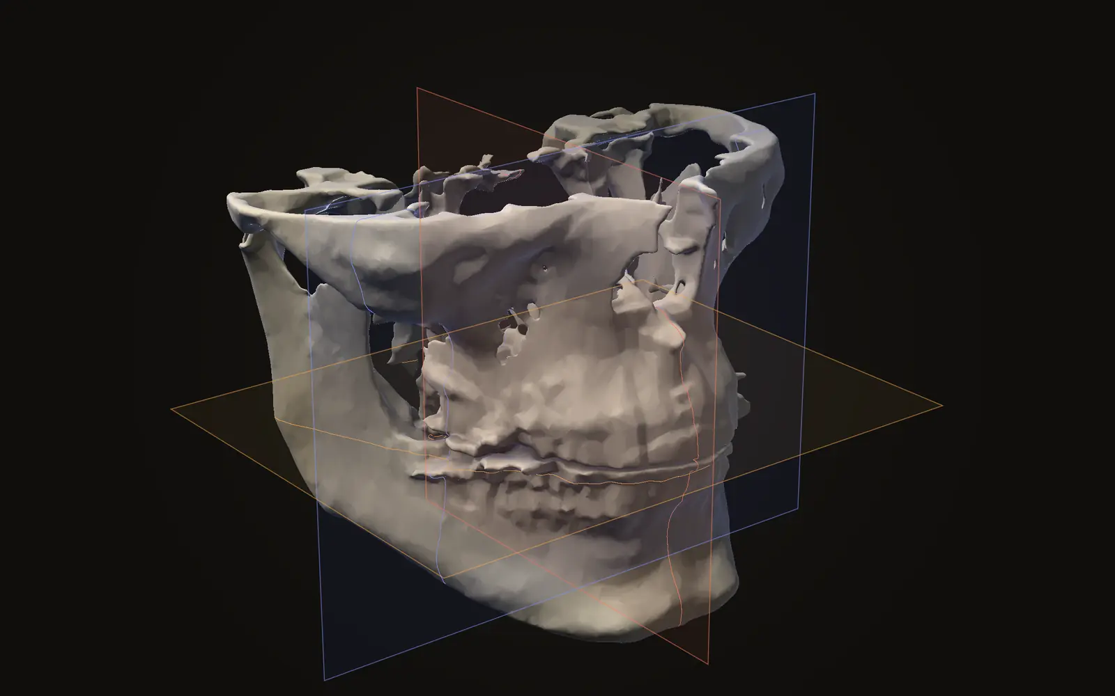
The same plane definition fixes the patient axes: x increases towards the patient's left, y towards the back, z towards the head.[4, C.7.6.2.1.1] The three standard views are cut along these axes, as axial, coronal and sagittal views explained shows.
Why can a header hold a name?#
DICOM began as a way to move image data between systems.[2] The data set can hold the patient's name as an attribute, next to the image attributes. The standard's confidentiality annex lists Patient's Name 0010,0010 among the attributes that a de-identification process deals with.[7, Annex E]
Treat a folder of scan files as a record with your name in it, whatever the files are called. A viewer that reads the files in your browser keeps that header on your computer, and the article on uploading a scan to a viewer website shows how to check that for any viewer. Jaw Viewer reads the files in the tab and has no upload step.
Why does a scan come as hundreds of files?#
Because each file holds one image.[1, §7] A scan of 400 slices would be 400 files. Each header carries its own slice position, which is what lets software sort the slices and stack them into a volume.[4, C.7.6.2]
Other medical formats, such as Analyze, NIfTI and MINC, are built differently from DICOM.[6]
The opening-your-scan guide lists what the viewer reads from the folder and what it warns about.
What is a DICOM file?
It is one medical image stored with a header of labelled attributes, such as scanner, image size and voxel size. A CBCT scan is usually hundreds of these files, one per slice.
Does a DICOM file contain the patient's name?
It can. The header has an attribute for the patient's name, and the standard lists it among the attributes to remove when a file is de-identified. Whether it is filled in depends on the clinic.
Why does my scan file have no .dcm ending?
The standard does not make a file name or extension part of the format. Software recognises a DICOM file by the letters DICM near its start, so I0000001 is a normal name.
Is a DICOM file the same as a JPEG?
No. A JPEG is a picture. A DICOM file holds the pixel values plus the details needed to turn a stack of slices into a measured 3D scan.
Sources
- DICOM Standards Committee (2026). DICOM PS3.10, Media Storage and File Format for Media Interchange, section 7 (DICOM File Format). NEMA. Version 2026d, read 21 November 2026.Link
- Mildenberger P, Eichelberg M, Martin E (2002). Introduction to the DICOM standard. Eur Radiol, 12: 920-927. Abstract read. Review of the history, concept and structure of DICOM.DOIPubMed
- DICOM Standards Committee (2026). DICOM PS3.5, Data Structures and Encoding, section 7 (The Data Set). NEMA. Version 2026d, read 21 November 2026.Link
- DICOM Standards Committee (2026). DICOM PS3.3, Information Object Definitions, section C.7.6.2 (Image Plane Module). NEMA. Version 2026d, read 21 November 2026.Link
- DICOM Standards Committee (2026). DICOM PS3.5, Data Structures and Encoding, section 8.2 (Native or Encapsulated Format Encoding). NEMA. Version 2026d, read 21 November 2026.Link
- Larobina M, Murino L (2014). Medical image file formats. J Digit Imaging, 27: 200-206. Abstract read. Compares Analyze, NIfTI, MINC and DICOM.DOIPubMed
- DICOM Standards Committee (2026). DICOM PS3.15, Security and System Management Profiles, Annex E (Attribute Confidentiality Profiles). NEMA. Version 2026d, read 21 November 2026.Link
Terms used
- DICOM
- The file format and standard that medical scanners use for images and their details.
- Series
- One set of images from a single acquisition. A CBCT scan is usually one series of hundreds of slices.
- Grey value
- The brightness number stored for a voxel. On a CBCT scan it is not a calibrated measure of density.
- Voxel
- A three-dimensional pixel. A scan is a grid of voxels, each with one grey value.
This is general information, not medical advice. Jaw Viewer is not a medical device. Talk to your surgeon, orthodontist or radiologist about your own case.