Key points
- A cephalometric analysis is a set of angles and distances calculated from named landmark points on a head image, so every result depends on where those points were placed.
- The Wits appraisal (Jacobson, 1975) drops perpendiculars from points A and B onto the occlusal plane and reads how far apart they land, instead of using a cranial base line.
- In Jacobson's sample of 21 men and 25 women with excellent occlusion, the two Wits points coincided on average in women and sat 1 mm apart in men.
- Three analyses applied to the same 88 cephalograms of children gave significantly different classifications on every parameter, so the method chosen changes the report.
A cephalometric analysis is a set of angles and distances calculated between named landmark points on a head image, traditionally a side-view X-ray called a cephalogram or head film.[1] The Steiner and Downs analyses and the Wits appraisal each pick different points and reference lines, so the same head can give different readings depending on which one a report uses. Every result depends on where someone placed the points.
Where do these analyses come from?#
Cephalometry is older than 3D scans. Downs published a paper on variations in facial relationships in 1948,[2] Steiner followed in 1953 with a paper titled "Cephalometrics for you and me",[3] and Jacobson described the Wits appraisal in 1975.[4] Their names now stand for sets of measurements. The descriptions below follow later papers that use the measurements, plus the 1975 abstract.
What do SNA, SNB and ANB measure?#
The Steiner analysis relies on the SN plane, a line between two cranial base landmarks, sella and nasion, and on the ANB angle.[5] The letters spell out the points. SNA is the angle sella-nasion-A point, and SNB is sella-nasion-B point.[5] Points A and B sit on the upper and lower jaw respectively.[4] SNA is read as the position of the upper jaw and SNB as the position of the lower jaw, both against the cranial base.[5] ANB, written as point A-nasion-point B,[5] compares the two jaws with each other.
Each angle has a weak spot: it rests on the SN plane. The 2025 study notes that Steiner's method assumes the cranial base landmarks are placed consistently.[5] If nasion is placed a little differently, SNA, SNB and ANB all move.
How is the Downs analysis different?#
Downs analysis typically uses the Frankfort horizontal and vertical references rather than the SN plane,[5] and its Y-axis is used to judge the direction of facial growth.[5] Steiner therefore uses a line through the skull base, while Downs uses the Frankfort lines.
What does the Wits appraisal add?#
Jacobson proposed a method that avoids the cranial base. You draw perpendiculars from point A on the upper jaw and point B on the lower jaw onto the occlusal plane, the plane along which the teeth meet. The two contact points are named AO and BO.[4] The result is how far apart AO and BO land along that plane.
Jacobson measured 21 men and 25 women selected for excellent occlusion. On average, AO and BO coincided in the women, and BO sat 1 mm ahead of AO in the men.[4] He reported that in a skeletal Class II pattern BO would sit well behind AO (a positive reading), and in Class III the reading would be negative.[4] He presented the appraisal as having advantages over the ANB reading.[4]
| Analysis | Reference it uses | What it reads |
|---|---|---|
| Steiner (1953) | SN plane[5] | SNA, SNB and ANB angles[5] |
| Downs (1948) | Frankfort horizontal and vertical[5] | Y-axis, used for growth direction[5] |
| Wits (1975) | Occlusal plane[4] | Distance between AO and BO[4] |
Do different analyses agree?#
Not always. A 2025 study traced 88 cephalograms of mouth-breathing children aged 6 to 14 with the Sassouni, Down and Steiner methods. It found significant differences between the three on every parameter it assessed, with Steiner showing more retrognathic jaws than Sassouni did.[5] The authors called this diagnostic inconsistency. It is one sample of children with one condition, but it shows why a report should say which analysis it used. The same paper also notes that differences in locating points such as nasion or gonion, or in head position, can influence angular measurements.[5]
What changes in 3D?#
On a CBCT scan the same kind of points are placed on a 3D scan instead of a flat film. A 2014 systematic review of 3D cephalometry kept 35 publications and found limited evidence for its diagnostic efficacy. Only 6 studies met the criteria for a moderate level of evidence, and the authors called for methodologically standardised studies.[6] How reliably points can be found is the subject of landmark placement error in cephalometry. For the wider limits of measuring on a scan, see how accurate CBCT measurements are.
Seeing the points on a scan#
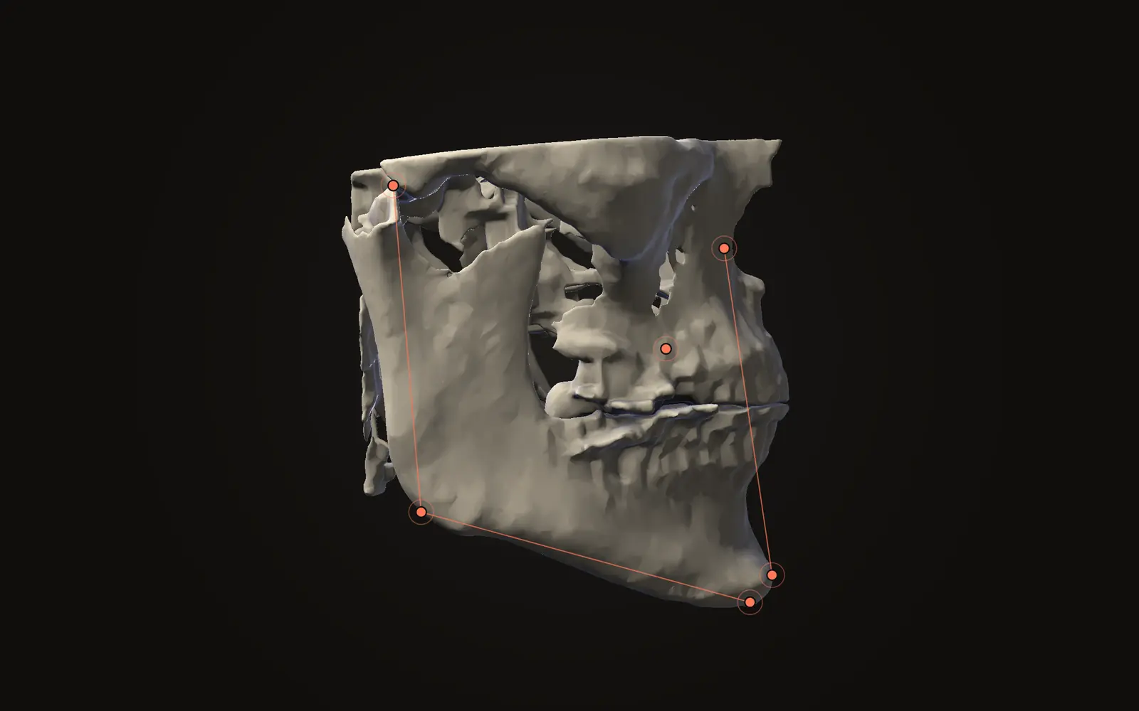
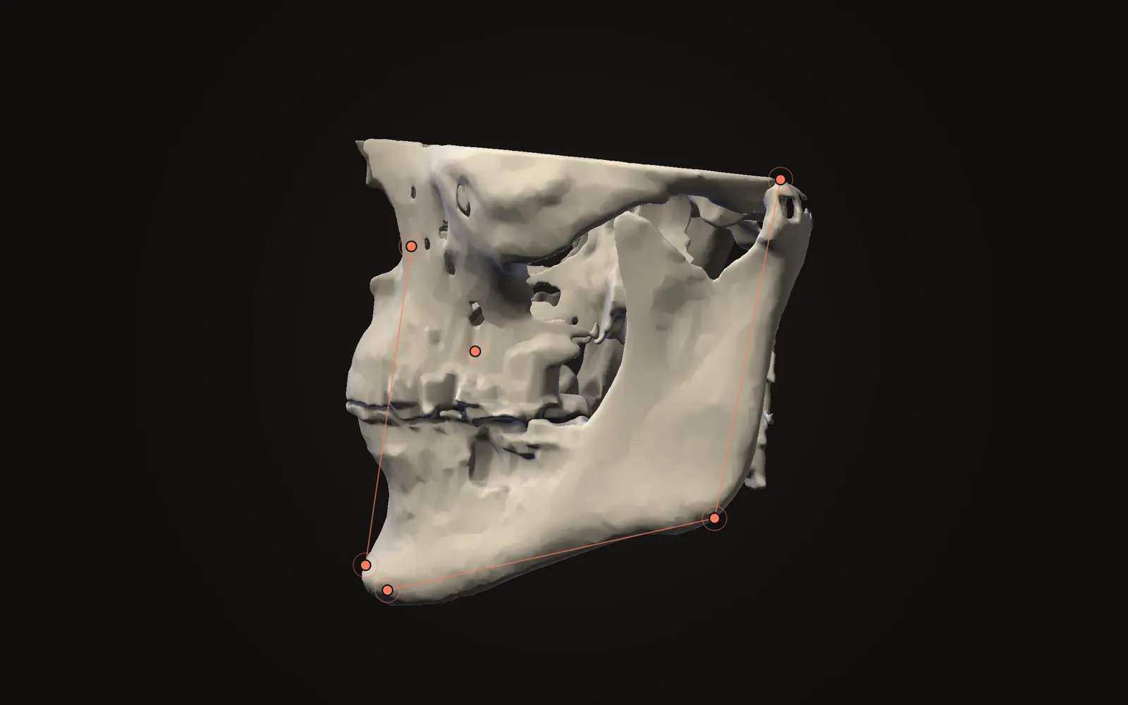
Jaw Viewer does not place anything for you. You choose a guided landmark set, read the prompt for the next point, and place it in the slices, checking it from all three directions before you go on. The analysis controls then list the calculations available for your landmark set, and a missing point has to be placed before its result appears. If an angle surprises you, look at the points behind it first. The landmarks and analysis guide has the steps, and how to measure on a CT scan covers distances and angles in general.
Use the numbers to understand the points and their relationships. A reference value is not a diagnosis and not a target, and a result tells you nothing until it is read with the clinician who knows the person. To see what the jaws look like and how Class II or Class III patterns come into jaw operations, read what orthognathic surgery is. For what the scan itself shows, see what a CBCT scan shows.
What is a cephalometric analysis?
It is a set of angles and distances calculated between named landmark points on a head image. The best known are the Steiner and Downs analyses and the Wits appraisal.
What do SNA, SNB and ANB mean?
SNA is the angle sella-nasion-A point, SNB is sella-nasion-B point, and ANB is point A-nasion-point B. They describe where the upper and lower jaw sit relative to the cranial base.
What is the Wits appraisal?
A method from 1975 that drops perpendiculars from points A and B onto the occlusal plane and measures how far apart the two contact points land. Positive and negative readings point to different jaw patterns.
Can I work out my diagnosis from these numbers?
No. A result depends on the points placed, the head position and the analysis chosen, and reference values come from specific groups. Take the numbers to your clinician with your questions.
Sources
- Baumrind S, Frantz RC (1971). The reliability of head film measurements. 1. Landmark identification. Am J Orthod, 60(2): 111-127. Cited for its title and topic only; no abstract is available.DOIPubMed
- Downs WB (1948). Variations in facial relationships; their significance in treatment and prognosis. Am J Orthod, 34(10): 812-840. Cited for its title and year only; no abstract is available.DOIPubMed
- Steiner CC (1953). Cephalometrics for you and me. Am J Orthod, 39(10): 729-755. Cited for its title and year only (Crossref record); no abstract is available.DOI
- Jacobson A (1975). The "Wits" appraisal of jaw disharmony. Am J Orthod, 67(2): 125-138. Read from the PubMed abstract.DOIPubMed
- Kumari B, Singh RK, Khanna R (2025). Cephalometric evaluation of mouth-breathing patients: a retrospective study using Sassouni, Down, and Steiner analyses. Cureus, 17: e97925. 88 cephalograms of mouth-breathing children aged 6 to 14. Open access.DOIPubMed
- Pittayapat P, Limchaichana-Bolstad N, Willems G, et al. (2014). Three-dimensional cephalometric analysis in orthodontics: a systematic review. Orthod Craniofac Res, 17(2): 69-91. 35 publications included.DOIPubMed
Terms used
- Cephalogram
- A standardised side-view X-ray of the head, also called a head film, used to place landmarks and measure angles between them.
- Occlusal plane
- The plane along which the upper and lower teeth meet when the mouth closes.
- CBCT
- Cone beam computed tomography. A scanner that rotates once around the head and records a cone-shaped X-ray beam, common in dentistry and jaw surgery.
This is general information, not medical advice. Jaw Viewer is not a medical device. Talk to your surgeon, orthodontist or radiologist about your own case.