Key points
- A 2019 systematic review of 13 CBCT studies found that landmarks on the midsagittal plane were identified more reliably than bilateral landmarks such as condylion, orbitale and porion.
- In one study the review tabulated, sella was placed with a spread of 0.50 mm (SD 0.24) across 9 observers, and point B with 1.50 mm (SD 0.72).
- The studies were too different to pool, so the review reports no single error figure for 3D landmarks, and a 2014 review found only 6 of 35 studies reached moderate evidence.
- Repeat placements by the same person usually agree better than placements by different people, so a number is most comparable with one made by the same observer.
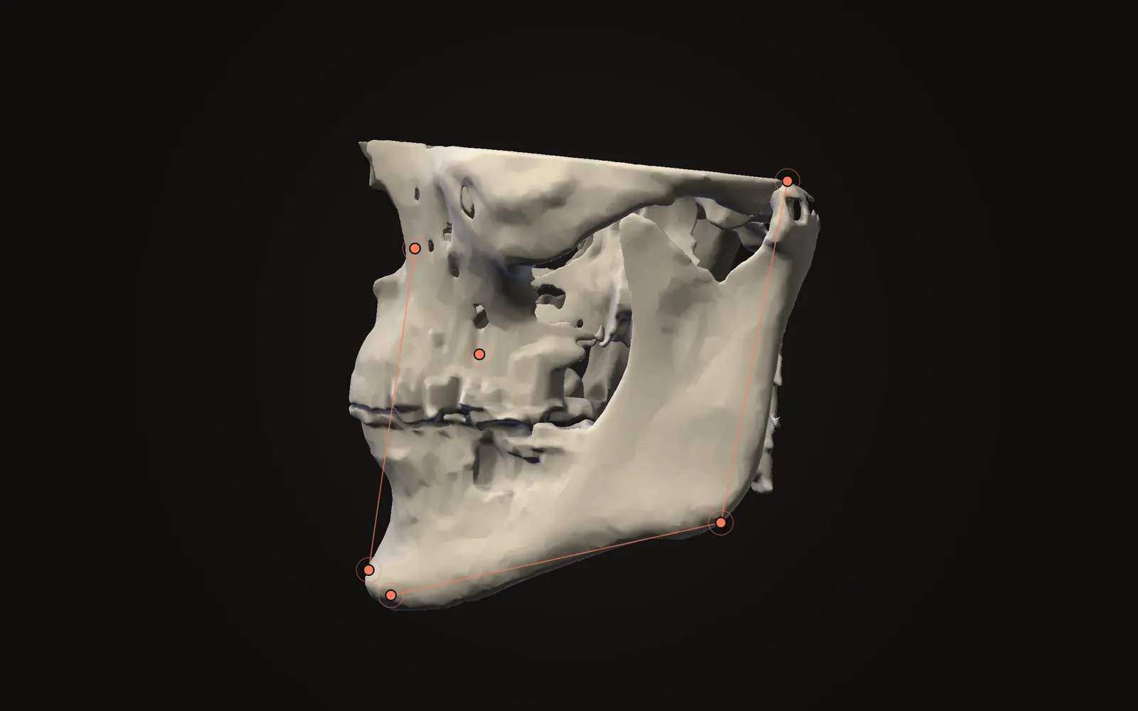
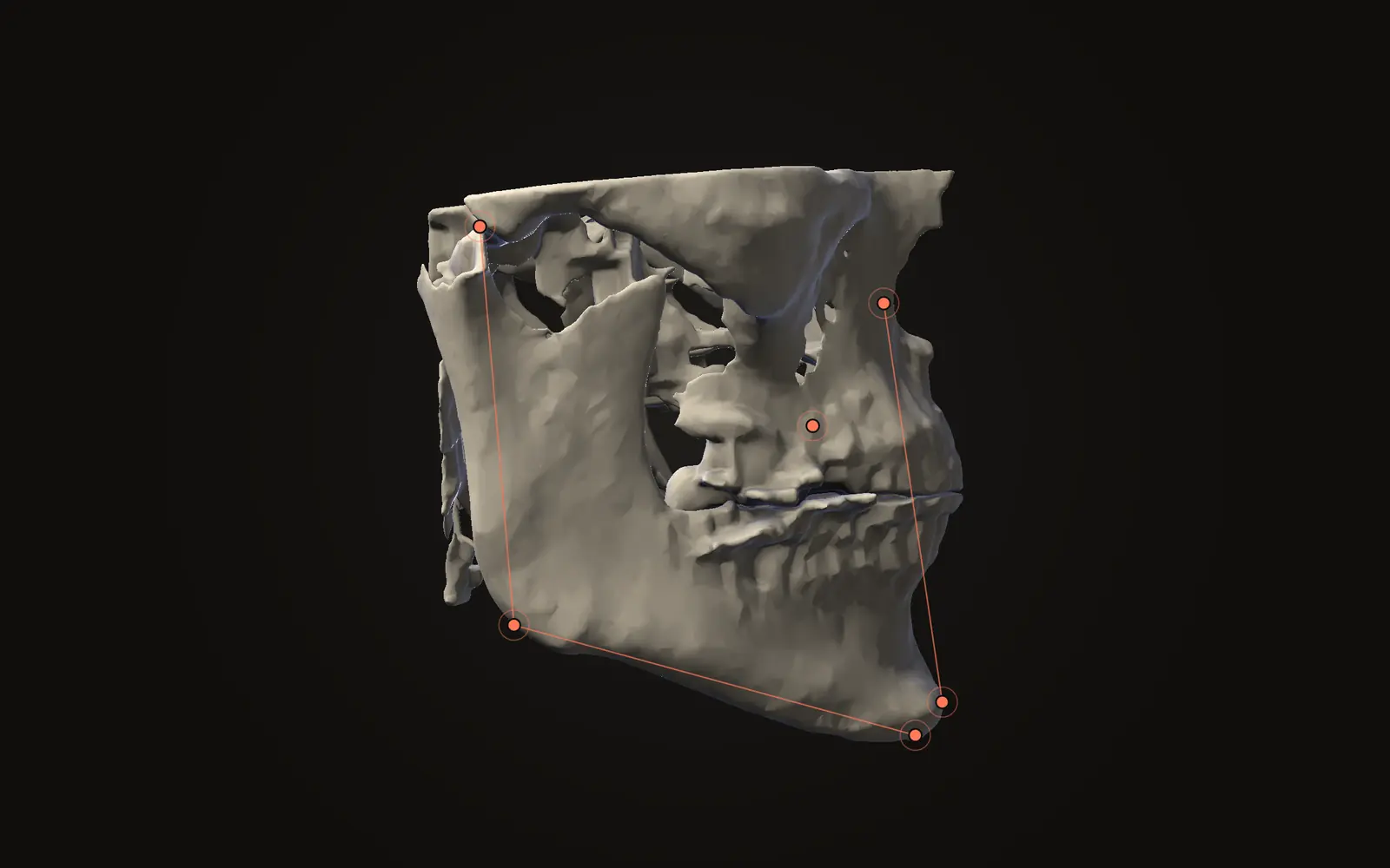
A cephalometric landmark is a named point on the skull, such as nasion or menton, and every angle in an analysis is built from several of them. Each point is placed by a person, and people do not put it in the same spot twice. A 2019 review of 13 CBCT studies found that points on the midsagittal plane, the plane that splits the head into a left and a right half, were identified more reliably than points that come in left and right pairs.[1]
Where the error comes from#
A landmark is a definition, such as "the most anterior point of the chin", applied to a picture. A sharp corner leaves little room for judgement; a long curve leaves much more. The 2019 review noted that landmarks along broad curvatures, or with indistinct boundaries, were harder to locate and showed more error.[1, Discussion]
The problem is old. Baumrind and Frantz studied landmark identification on lateral head films in 1971, long before CBCT.[2] The 2019 review lists projection errors among the limits of the flat film that CBCT may overcome.[1] A three-dimensional scan still leaves the judgement about where a point lies.
Same person or different people?#
Studies measure two kinds of disagreement. Intraobserver error is how far one person's repeated placements spread. Interobserver error is how far different people's placements spread. In the CBCT studies the 2019 review gathered, repeat placements by the same examiner were typically more reliable than comparisons between examiners.[1, Results of individual studies]
Two angles measured by the same person on two scans share much of the same placement habit. Measurements by different people carry both sets of habits.
Which landmarks hold steady?#
The review sorted landmarks by how consistently they were found. Those it recommended for 3D work, on the midsagittal plane, were sella, basion, nasion, anterior nasal spine, A point, B point, pogonion, gnathion and menton.[1, Discussion] Bilateral landmarks with variable consistency included those on the condyles, orbitale, porion and lingula.[1, Discussion]
The review suggests a reason: points along the midline are easy to find because the sagittal slice looks much like a lateral cephalogram.[1, Discussion] A pair of points, in contrast, has to be found twice, on two sides, in a head that may be slightly turned.
What the numbers looked like#
The review could not pool its studies, because they differed in design, observers and landmark lists, so it gives no single error figure.[1] Its tables do list figures from individual studies, which show the range.
| Landmark | Reported spread between observers | Study (as tabulated in the review) |
|---|---|---|
| Sella | 0.50 mm (SD 0.24) | 19 scans, 9 orthodontic residents[1, Tables 3 and 11] |
| Point B | 1.50 mm (SD 0.72) | 19 scans, 9 orthodontic residents[1, Tables 3 and 11] |
| Orbitale | 5.76 mm, mediolateral direction | 20 scans, 5 observers[1, Tables 3 and 14] |
| Porion | 7.14 mm, mediolateral direction | 20 scans, 5 observers[1, Tables 3 and 14] |
The two studies measured spread in different ways, so compare the rows within a study, not between them. In the first study, the average across 32 landmarks was 1.64 mm.[1, Table 3]
How a small error becomes an angle#
Take a line 60 mm long between two landmarks, an illustrative length. If one end moves 1.5 mm sideways, the line turns by about 1.4 degrees (our arithmetic). An angle built from three or four points can collect several such shifts, in either direction. Short lines turn further for the same shift, which is why angles that depend on closely spaced points are more sensitive to placement.
This is also why an analysis result should be read together with the points behind it. What a cephalometric analysis is walks through how common analyses draw on those points.
Does 3D make this go away?#
A 2014 review of 3D cephalometry looked at measurement accuracy and the reproducibility of landmark identification. It included 35 publications and found limited evidence for diagnostic efficacy. Only 6 studies met the criteria for moderate evidence, and the authors called for standardised studies.[3]
One CBCT study in the 2019 review compared two ways of looking at the same scans, slices against a 3D rendering. It found landmark identification more reliable in the slices.[1, Table 3] That finding comes from a single study of 12 scans and 3 observers. It supports checking a point in more than one view, though the evidence is limited.
Placing landmarks in the viewer#
In Jaw Viewer, every landmark is placed by you, one at a time, in a guided set that prompts for the next point. Check each point in the slices from all three directions before moving on, because a point that looks right in one view can be off in another. The landmarks guide lists the steps, and the limits page says what a measurement depends on.
A good exercise is to place a set, note the angles the analysis lists, and place the set again another day without looking at the first result. The difference between the two runs is your own intraobserver error for that scan. How far point placement matters for plain distances is covered in how accurate CBCT measurements are, and the steps for a distance are in how to measure on a CT scan. If you compare two dates, landmark-based alignment brings the same issue into the comparison, covered in registration error when comparing two scans.
What is landmark identification error?
It is the difference between where a point is placed and where it was placed before, by the same person or by another. Every angle and distance built from that point inherits the difference.
Which cephalometric landmarks are most reliable on CBCT?
A 2019 review found landmarks on the midsagittal plane, such as sella, nasion, A point, B point and menton, more reliable than bilateral ones such as porion, orbitale and the condyles.
Is landmark error smaller in 3D than on a 2D cephalogram?
The evidence is mixed. A 2019 review of 13 CBCT studies could not pool its data, and a 2014 review found only 6 of 35 studies reached a moderate level of evidence.
Why does the same person get different results on different days?
Many points sit on curved bone or soft edges with no sharp boundary, so the exact spot is a judgement. Studies repeat placements at intervals, such as one week, to measure that spread.
Sources
- Sam A, Currie K, Oh H, et al. (2019). Reliability of different three-dimensional cephalometric landmarks in cone-beam computed tomography: A systematic review. Angle Orthod, 89(2): 317-332. 13 studies of human CBCT scans, mostly moderate quality, too varied to pool. Tables read in the free full text (PMC8120873).DOIPubMed
- Baumrind S, Frantz RC (1971). The reliability of head film measurements. 1. Landmark identification. Am J Orthod, 60(2): 111-127. Cited for its topic only; PubMed has no abstract for it.DOIPubMed
- Pittayapat P, Limchaichana-Bolstad N, Willems G, et al. (2014). Three-dimensional cephalometric analysis in orthodontics: a systematic review. Orthod Craniofac Res, 17(2): 69-91. 35 publications included; searched to March 2012.DOIPubMed
Terms used
- Intraobserver error
- The spread between repeated placements of the same point by the same person on different occasions.
- Interobserver error
- The spread between placements of the same point by different people.
This is general information, not medical advice. Jaw Viewer is not a medical device. Talk to your surgeon, orthodontist or radiologist about your own case.