Key points
- The mandibular canal starts at the mandibular foramen on the inner side of the ramus, runs down and forward, then horizontally through the body of the lower jaw to the mental foramen.
- A 2010 review quotes a canal diameter of 2.0 to 2.4 mm near the third molar in a study of 45 Asian adults, so on a scan the canal is only a few voxels wide.
- A 2022 meta-analysis of 16 CBCT studies (8,862 canals) found a bifid canal in about 38.0%, an accessory mental foramen in 9.54% and a retromolar foramen in 23.64%.
- Bifid canal figures depend on the method: 0.08% to 0.95% in four panoramic radiograph studies listed in a 2010 review, and 65% of patients in one study that reconstructed images in many planes.
The inferior alveolar nerve canal, also called the mandibular canal, is a channel inside the lower jaw. It starts on the inner surface of the ramus, the upright part of the jaw, and runs forward to an opening near the chin. It carries the inferior alveolar nerve and the blood vessels beside it.[1] On a CBCT scan you follow it through the slices, and its position and shape differ from person to person.
Where does the canal run?#
The canal begins at the mandibular foramen on the medial (inner) surface of the ascending ramus. It runs obliquely downward and forward in the ramus, then horizontally forward through the body of the lower jaw until the mental foramen. Beyond that opening, the continuation is called the incisive canal.[1]
Along the way the canal also moves from side to side. A review reports that it usually crosses from the lingual (tongue) side towards the buccal (cheek) side, and that in most cases it sits midway between the two outer plates of bone near the first molar.[1] In the molar region the nerve divides into the mental and the mandibular incisal nerves.[1]
The canal is narrow. One study of 45 Asian adults, quoted in the same review, found that near the third molar the canal is usually a single structure 2.0 to 2.4 mm in diameter. Cadaver studies in the review give other widths, so there is no single figure.[1] A scan with 0.4 mm voxels would put about five or six voxels across a canal of that size, which is why the voxel size of your scan limits how sharply the wall can be seen.
What does it look like on a scan?#
The 2010 review describes the canal on panoramic radiographs as a dark ribbon of radiolucency flanked by two radiopaque white lines.[1] On a CBCT scan you look for the same dark channel inside the bone, in slices instead of a flat image. A single axial slice cuts across the canal at one spot, so it helps to follow it along the jaw in the sagittal and cross-sectional views too. The three standard directions are explained in axial, coronal and sagittal views.
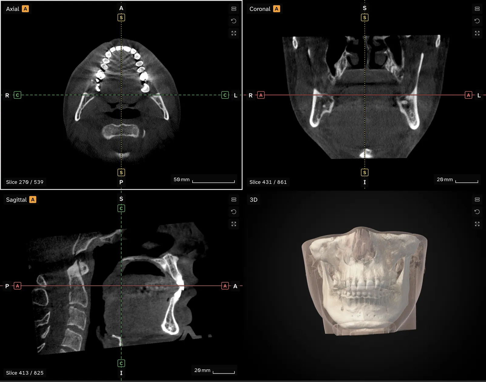
Brightness and contrast change what you see at the edge of the canal, and the grey values on a CBCT scan are not calibrated densities. What a CBCT scan shows covers why.
How much does the canal vary?#
A 2010 literature review of 46 sources concluded that the canal and its bundle vary with the individual, sex, age, race, the assessment technique and the degree of bone loss in a toothless ridge.[1] It describes several canal shapes in the vertical plane. The canal may run lower as it goes forward, have a sharp decline, or hang in a catenary-like curve.[1]
In a study of panoramic radiographs described there, a canal counted as high if it lay within 2 mm of the root tips of the first and second molars.[1] That is a way of sorting canals by height in one study, and it is not a target for anyone's scan.
What do the extra branches and openings add up to?#
A 2022 systematic review pooled 16 cross-sectional CBCT studies covering 8,862 canals. It counted branches and extra openings that leave the main canal:[2]
| Feature | Incidence reported across studies |
|---|---|
| Accessory mental foramen | 9.54% (95% CI 6.39 to 12.69) |
| Retromolar foramen | 23.64% (95% CI 14.44 to 32.84) |
| Bifid mandibular canal | about 38.0% |
| Mandibular lingual foramina | found in almost all adults studied |
These are pooled figures from many groups of people. They say how often such features were reported in research, and they are not an expectation for any one scan. The review's authors describe significant anatomical variation among individuals.[2] What each feature is, and how to follow the canal to its exit at the chin, is the subject of the article on the mental foramen and canal variations.
Why do bifid canal figures differ so much?#
The imaging method affects what is found. The 2010 review lists four studies of panoramic radiographs with bifid canal figures of 0.9%, 0.08%, 0.95% and 0.35%. It also describes a study that reconstructed 122 images in various planes through the ramus and saw a bifid canal in 65% of patients and 43% of sides.[1] The review takes this as a sign of how much the diagnostic method matters.[1]
How to trace the canal in the viewer#
The panoramic view guide sets out the steps. Jaw Viewer follows a curve you set along the dental arch and draws a panoramic reconstruction from the scan, with a strip of cross-sections beside it. The reconstruction adds no detail the scan did not record, and a misplaced curve gives a misleading picture, as panoramic reconstruction from CBCT explains.
The canal tool lets you mark the path of the canal by hand and shows that path in the other views. The line is an estimate of where the canal runs, and it is only as good as the points you place. Check it in the cross-sections instead of trusting a smooth 3D tube. A canal that is faint in one section may be clear two sections along, and a line that looks fine in 3D can leave the canal in the slices.
Placing points by hand has the same limits as any other measurement on a scan. How accurate are measurements on a CBCT scan covers them, and the limits page says what a number on screen can and cannot tell you. The viewer shows where the canal appears to run in your images. It says nothing about how the nerve works.
The canal also matters in lower jaw operations, because the nerve runs through the bone that is cut. BSSO, the lower jaw split operation, covers what studies report about lip sensation after one.
Where does the inferior alveolar nerve canal run?
It begins at the mandibular foramen on the inner surface of the ramus, runs obliquely down and forward, then horizontally through the body of the lower jaw to the mental foramen.
How wide is the mandibular canal?
One study of 45 Asian adults, quoted in a 2010 review, found a single canal of 2.0 to 2.4 mm diameter near the third molar. Other studies in the review give other widths.
How common is a bifid mandibular canal?
A meta-analysis of 16 CBCT studies reported about 38.0%. Panoramic radiograph studies listed in a 2010 review gave 0.08% to 0.95%. The method used to look decides much of the figure.
Does Jaw Viewer find the nerve canal by itself?
No. You mark the path of the canal by hand and check it in the cross-sections. The line is your estimate of where the canal runs, not a detected structure.
Sources
- Juodzbalys G, Wang HL, Sabalys G (2010). Anatomy of mandibular vital structures. Part I: mandibular canal and inferior alveolar neurovascular bundle in relation with dental implantology. J Oral Maxillofac Res, 1(1): e2. Literature review of 46 sources, searched up to November 2009.DOIPubMed
- Shan S, Zhong S, Li J, et al. (2022). Systematic review and meta-analysis of mandibular canal variations on cone beam computed tomography. Oral Radiol, 38(4): 445-451. 16 cross-sectional CBCT studies, 8,862 canals.DOIPubMed
Terms used
- Mandibular canal
- The channel inside the lower jaw that carries the inferior alveolar nerve and blood vessels. It starts on the inner surface of the ramus and ends at the mental foramen.
- Inferior alveolar nerve
- The nerve that runs through a canal inside the lower jaw and supplies feeling to the lower teeth, lip and chin.
- Mental foramen
- The opening on the outer surface of the lower jaw where the canal ends and the mental nerve comes out.
This is general information, not medical advice. Jaw Viewer is not a medical device. Talk to your surgeon, orthodontist or radiologist about your own case.