Key points
- A 2018 systematic review of 22 studies (2 clinical, 20 ex vivo) found a wide range of error in CBCT linear measurements, with both over- and underestimation against a gold standard.
- A 2024 meta-analysis of low-dose protocols pooled a measurement error of -0.24 mm (95% CI -0.52 to 0.04), a tendency to read slightly short, with high heterogeneity.
- In 28 dry skulls, average error on easily seen orthodontic wires was under 0.6% in axial slices and under 1.2% on panoramic reconstructions; anatomical points varied more.
- The authors of the 2018 review suggest a 2 mm safety margin to neighbouring structures, because motion, metal and software can lower accuracy in practice.
Studies of CBCT linear measurements report a wide range of error, and the error runs both ways: some distances come out too long, some too short, compared with a physical gold standard.[1] On easily seen test wires the average error was small, and on anatomical points it was larger and more variable.[2] The number on screen is only as good as the scan and the points placed on it.
What the reviews looked at#
A 2018 systematic review set out to summarise how accurate linear CBCT measurements are for implant work, such as bone height, bone width and the distance to a nearby structure. After 2,516 titles it kept 22 studies. Two were clinical and 20 were ex vivo, meaning measured on specimens such as skulls.[1] The reviewers compared each measurement with a gold standard. They found high accuracy and reliability on cross-sectional images, together with a wide range of error and both over- and underestimation.[1]
A 2024 review looked only at low-dose protocols, which use less exposure to keep dose down. It included 13 studies (81 samples in total), nine of them pooled in a meta-analysis. The pooled measurement error was -0.24 mm (95% CI -0.52 to 0.04), with high heterogeneity, and it showed a tendency to underestimate real values.[3] A negative error means the scan read slightly shorter than the real distance. The confidence interval reaches just past zero, so the review cannot say the shift is certain.
22 studies
in the 2018 review: 2 clinical, 20 ex vivo[1]
-0.24 mm
pooled error, low-dose protocols (95% CI -0.52 to 0.04)[3]
0.3 to 0.4 mm
voxel size judged adequate for implant planning images[1]
Which numbers are real patients?#
Most studies used lab specimens. Of the 22 studies in the 2018 review, only two involved patients.[1] The 2024 review used a tool for in vitro studies to judge bias, so its studies were lab work as well.[3]
The 2018 authors write that clinical accuracy and reliability are most likely reduced by patient motion, metallic artefacts, device-specific exposure settings, the software used, and manual versus automated procedures.[1] The review figures are best read as a best case, not a promise for any one scan.
Voxel size and sharpness#
A voxel is the smallest cell of the scan, and its size sets how finely a point can be placed. The 2018 review concluded that a voxel size of 0.3 to 0.4 mm gives images of acceptable diagnostic quality for implant treatment planning.[1] The 2024 review found that several studies saw no significant difference between protocols that differed in voxel size, current (mA) or dose level. Some did note exceptions in the posterior mandible.[3] So a smaller voxel does not guarantee a better number. For how voxel size relates to sharpness, see voxel size and sharpness on CBCT.
Does the head position change it?#
One skull study checked this directly. Ludlow and colleagues scanned 28 dry skulls in an ideal position, shifted, and rotated, then compared software measurements with caliper measurements. Skull position and measuring the right or left side did not significantly change accuracy.[2]
They also measured in two ways: on panoramic reconstructions (two-dimensional) and directly on axial slices (three-dimensional). For easily visualised orthodontic wires, the average error was under 1.2% with the two-dimensional technique and under 0.6% with the three-dimensional one.[2] On a 50 mm distance, 0.6% is 0.3 mm (our arithmetic). But the same study found that measurements between anatomical points were significantly more variable, whichever technique was used.[2]
| Measurement | Result | Source |
|---|---|---|
| Linear measurements, 22 studies | Wide error range, over and under | [1] |
| Low-dose protocols, pooled error | -0.24 mm (95% CI -0.52 to 0.04) | [3] |
| Wires, axial slices | Average error under 0.6% | [2] |
| Wires, panoramic reconstruction | Average error under 1.2% | [2] |
| Anatomical points | Significantly more variable | [2] |
Placing the point#
A wire is an obvious target. A point on bone is a judgement about where an edge or a centre lies, and the study reports only that these measurements varied more, not why. How far hand-placed points disagree is covered in landmark placement error in cephalometry.
Measuring in the viewer#
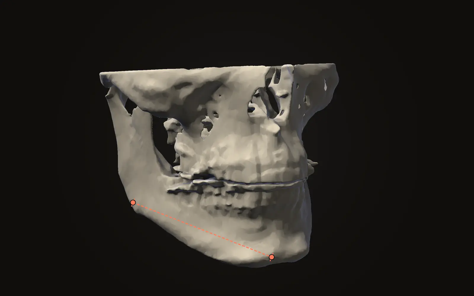
In Jaw Viewer, a distance is two points, shown in millimetres, and the same line appears in the slices and on the bone. The reading depends on where you put the points, so a line placed on a sharp edge is steadier than one placed on a soft-looking boundary. The measuring guide lists the tools, and the limits page says what a measurement here depends on. For the steps, see how to measure on a CT scan. For how a cephalometric reading is built from points, see what a cephalometric analysis is.
If you compare two dates, the line-up adds its own error, covered in registration error when comparing two scans. For what the scan can and cannot show, see what a CBCT scan shows.
How accurate are CBCT measurements?
Reviews report a wide range of error, with both over- and underestimation. In 28 dry skulls, easily seen wires measured within 0.6% in axial slices. Points on bone varied more.
Does a smaller voxel size make a measurement more accurate?
Not by itself. One review found 0.3 to 0.4 mm voxels adequate for implant planning images, and a meta-analysis found no significant differences between several protocols. Point placement and artefacts matter too.
Can CBCT measurements be trusted for real patients?
Most of the evidence comes from skulls and other lab specimens, not living patients. Of 22 studies in one review, 2 were clinical. Motion and metal can reduce accuracy in practice.
What is a 2 mm safety margin?
It is the margin the authors of a 2018 review say should be kept to neighbouring anatomical structures when using CBCT measurements. It is their statement about the evidence, not advice for any one scan.
Sources
- Fokas G, Vaughn VM, Scarfe WC, et al. (2018). Accuracy of linear measurements on CBCT images related to presurgical implant treatment planning: A systematic review. Clin Oral Implants Res, 29 Suppl 16: 393-415. 22 studies, 2 clinical and 20 ex vivo.DOIPubMed
- Ludlow JB, Laster WS, See M, et al. (2007). Accuracy of measurements of mandibular anatomy in cone beam computed tomography images. Oral Surg Oral Med Oral Pathol Oral Radiol Endod, 103(4): 534-542. 28 dry skulls compared with caliper measurements.DOIPubMed
- Carneiro ALE, Reis INR, Bitencourt FV, et al. (2024). Accuracy of linear measurements for implant planning based on low-dose cone beam CT protocols: a systematic review and meta-analysis. Dentomaxillofac Radiol, 53(4): 207-221. 13 studies, 9 in the meta-analysis; the studies are in vitro.DOIPubMed
Terms used
- CBCT
- Cone beam computed tomography. A scanner that rotates once around the head and records a cone-shaped X-ray beam, common in dentistry and jaw surgery.
- Voxel
- A three-dimensional pixel. A scan is a grid of voxels, each with one grey value.
This is general information, not medical advice. Jaw Viewer is not a medical device. Talk to your surgeon, orthodontist or radiologist about your own case.