Key points
- Registration lines up two scans of one person on a reference region both contain, such as the anterior cranial base, so that a difference elsewhere can be read as change.
- In 16 pre and post treatment CBCT pairs, voxel-based registration left a mean distance of 0.2 to 0.37 mm on the anterior cranial base and 0.2 to 0.45 mm on the zygomatic arch.
- A 2022 review of 13 studies reported translational accuracy of 0.05 to 1.76 mm and rotational accuracy of 0.10 to 1.09 degrees, so the error depends on the method and the study.
- The reference region changes the result: in 16 growing subjects one popular mandibular mask worked in only 2 of 16, and a mandibular body mask gave the most consistent results.
Registration is the step that lines up two scans of the same person so that the same bone sits in the same place before you compare them. It is judged on a reference region both scans contain, and some gap always remains. In one study of 16 pre and post treatment CBCT pairs, that gap averaged 0.2 to 0.45 mm on bone that should not have changed.[1]
What does registration do?#
Two scans never start aligned. The head sat a little differently in the scanner, and the scanned region may not cover exactly the same area. Registration finds the move and turn that bring one scan onto the other. You pick a reference region that both scans show, and the software matches that region.
Everything outside the reference region is then read relative to it. If the match is good, a lower jaw that sits in a different place on the second date shows up as a difference. If the match is poor, bone that never moved looks as if it did. For this reason the reference has to be a structure that stays put between the two dates. One study used the anterior cranial base, in the front part of the skull base, and the zygomatic arch (cheekbone).[1]
Three ways to line up scans#
A review of superimposition methods sorted them into three groups: landmark-based (matching hand-placed points), surface-based (matching 3D surface models) and voxel-based (matching the scan's own voxel values in a chosen region).[2] Of its 15 included studies, eleven used voxel-based registration, one used landmarks, and three compared several techniques.[2]
The review concluded that certain voxel-based and surface-based methods seem to work properly and outperform landmark-based methods. Most studies had methodological shortcomings: 12 of the 15 had a high risk of bias, and the authors call for further validation.[2] A second review, of 13 studies on jaw surgery outcomes, also found voxel-based protocols had the highest accuracy and reproducibility.[3]
How big is the leftover error?#
One study measured 16 adult patients with jaw discrepancies, each with a scan before and after treatment. The scans were registered on the anterior cranial base, then the mean distance between the two 3D models was measured at four regions. It ranged from 0.2 to 0.37 mm (SD 0.08 to 0.16) for the cranial base registration. Registering on the left zygomatic arch instead gave 0.2 to 0.45 mm (SD 0.09 to 0.27).[1]
The authors judged both to be accurate and reproducible. They also note that the zygomatic arch can serve as the reference when a smaller field of view leaves out the cranial base.[1] Our reading: where nothing moved, the true distance would be zero, so the 0.2 to 0.45 mm left over gives a sense of what registration adds to a comparison, in that sample and with that method.
0.2 to 0.37 mm
mean distance after registering on the anterior cranial base, 16 pairs[1]
0.2 to 0.45 mm
mean distance after registering on the zygomatic arch, 16 pairs[1]
0.05 to 1.76 mm
translational accuracy across methods in a 2022 review of 13 studies[3]
That review also gave a rotational accuracy of 0.10 to 1.09 degrees and a reproducibility error of 0.01 to 0.26 mm, with run times from a few seconds to 40 minutes.[3] The spread, 0.05 to 1.76 mm, is wide. A figure from one method on one sample is not a promise for the next pair of scans.
Does the reference region matter?#
Yes, and it can decide whether the result is usable. A study of 16 growing subjects compared three reference regions for registering the lower jaw. The first, the Björk mask, worked properly in only 2 of the 16 patients. Of the other two masks, the mandibular body mask (the jaw without teeth, alveolar bone, rami and condyles) gave the most consistent results.[4]
The subjects were still growing, so this is not a ready match for an adult comparison. The same two scans can give different answers depending on which region the software matches.
| Choice | What the research found |
|---|---|
| Method | Voxel-based and some surface-based methods seem better than landmark-based, but 12 of 15 studies had high risk of bias[2] |
| Reference on the skull | Cranial base 0.2 to 0.37 mm, zygomatic arch 0.2 to 0.45 mm mean distance[1] |
| Reference on the lower jaw | One mask worked in 2 of 16; the mandibular body mask was most consistent[4] |
Comparing two dates in Jaw Viewer#
Jaw Viewer lines up two scans with automatic alignment on the skull base. You can limit the region to anatomy both scans share, match point pairs by hand, or nudge the alignment yourself. The alignment only turns and shifts one scan to match the other; it never bends the images to agree. The compare guide walks through the steps.
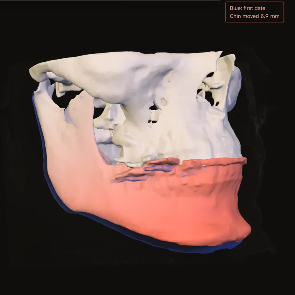
You can then view the pair side by side, blended, with a swipe, as a checkerboard, flickering between dates, as a difference, or as a distance map in 3D. The first thing to check is the reference region, in several slices and in an overlay. Teeth that line up do not prove the skull base does. Jaw Viewer gives no accuracy figure for its alignment, and the limits page says what the match depends on: missing anatomy, metal and different scan coverage all affect it.
A comparison also carries the usual measurement error of each scan. How far hand-placed points disagree is covered in landmark placement error in cephalometry, and the range of CBCT measurement error in how accurate CBCT measurements are. For what each scan can show on its own, see what a CBCT scan shows.
What is registration when comparing two CBCT scans?
It is lining up two scans of the same person so the same anatomy sits in the same place. You then compare what differs. The match is judged on a reference region both scans contain.
How accurate is voxel-based registration of CBCT scans?
In one study of 16 pre and post treatment pairs, the mean leftover distance was 0.2 to 0.45 mm on stable reference bone. A 2022 review reported translational accuracy from 0.05 to 1.76 mm across methods.
Why does the reference region matter?
The scans are matched on that region, so anything else is read relative to it. In one study of growing subjects, one mandibular mask worked in only 2 of 16 patients while another was more consistent.
Can a small difference between two scans be a real change?
A difference close to the registration error cannot be told apart from a slight misalignment. Research figures for that error come from specific methods and samples, so they do not transfer to a single comparison.
Sources
- Nada RM, Maal TJ, Breuning KH, et al. (2011). Accuracy and reproducibility of voxel based superimposition of cone beam computed tomography models on the anterior cranial base and the zygomatic arches. PLoS One, 6(2): e16520. 16 pre and post treatment CBCT pairs from adult patients with jaw discrepancies.DOIPubMed
- Mai DD, Stucki S, Gkantidis N (2020). Assessment of methods used for 3-dimensional superimposition of craniofacial skeletal structures: a systematic review. PeerJ, 8: e9263. 15 eligible studies out of 832.DOIPubMed
- Andriola FO, Haas Junior OL, Guijarro-Martínez R, et al. (2022). Computed tomography imaging superimposition protocols to assess outcomes in orthognathic surgery: a systematic review with comprehensive recommendations. Dentomaxillofac Radiol, 51(3): 20210340. 13 studies, 10 of them reporting accuracy.DOIPubMed
- Ruellas AC, Yatabe MS, Souki BQ, et al. (2016). 3D mandibular superimposition: comparison of regions of reference for voxel-based registration. PLoS One, 11(6): e0157625. 16 growing subjects, scans before and after treatment.DOIPubMed
Terms used
- CBCT
- Cone beam computed tomography. A scanner that rotates once around the head and records a cone-shaped X-ray beam, common in dentistry and jaw surgery.
- Registration
- Lining up two scans of the same person so the same anatomy sits in the same place, before you compare them. The match is judged on a reference region both scans share.
This is general information, not medical advice. Jaw Viewer is not a medical device. Talk to your surgeon, orthodontist or radiologist about your own case.