Key points
- Before and after jaw surgery scans are lined up (superimposed) on the cranial base, because that part of the skull is not changed by the operation. What differs elsewhere can then be shown as a coloured 3D map.
- In 10 patients scanned before and 1 week after upper jaw surgery, the average displacement over the lower jaw ramus surfaces was 0.77 mm, close to the 0.60 mm image resolution.
- Voxel-based superimposition on the anterior cranial base took 10 to 15 s in one study of 18 patients, with a superimposition error under 0.5 mm. The older open-source route takes about 3 hours.
- Jaw Viewer lines up two dates automatically on the skull base, then shows them side by side, blended, swiped, as a checkerboard, flickering, as a difference or as a distance map. It gives no accuracy figure.
Before and after jaw surgery scans are compared by lining them up on bone the operation does not change, the cranial base, and then looking at what differs elsewhere. The lining up is called superimposition or registration. In one study of CBCT scans taken before and 1 week after upper jaw surgery, the average displacement over the surfaces of the lower jaw's rami was 0.77 mm.[1]
Why not just lay one scan over the other?#
The head never sits in the scanner the same way twice. If you overlaid two scans as they come, the whole face would look as if it had moved. So one scan is turned and shifted until a region that should be the same in both lines up, and only then is the rest compared.
The reference has to be a region the operation leaves alone. In one study of 31 patients with a scan before and after surgery, the authors chose surgical patients because the reference area for the line-up was not affected by growth or by the surgical procedures.[2] That is why the cranial base, the bone behind and above the eyes and nose, is the usual choice for this kind of pair. The jaws and teeth moved, so matching on them would hide the very change you want to see.
Three ways to line up scans#
The literature describes three families of method: landmark-based, surface-based and voxel-based. Landmark-based means matching points picked on both scans. Surface-based matches 3D models of the bone. A voxel-based method compares the grey values of the scans themselves. All three use stable anatomical structures as the reference.[3]
Voxel-based is the one most often described, and the only one that can be fully automated.[3] A 2022 review of 13 studies on CT superimposition after jaw surgery found 7 that proposed or evaluated voxel-based methods and 3 that focused on surface-based ones.[4] The same review gives accuracy figures for the methods it covers, and the article on registration error goes through them and through what the leftover gap means.
10 to 15 s
fast voxel-based superimposition on the anterior cranial base, 18 patients[5]
under 5 minutes
commercial voxel-based method, 31 surgical patients[2]
about 3 hours
the older open-source method it was compared with[2]
What do the two scans show once they are lined up?#
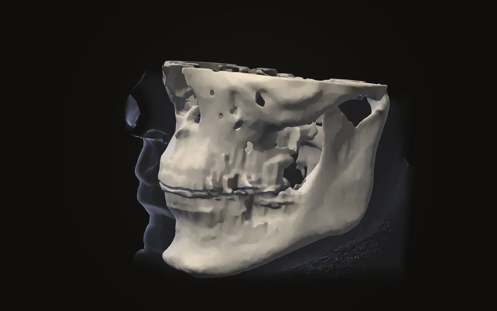
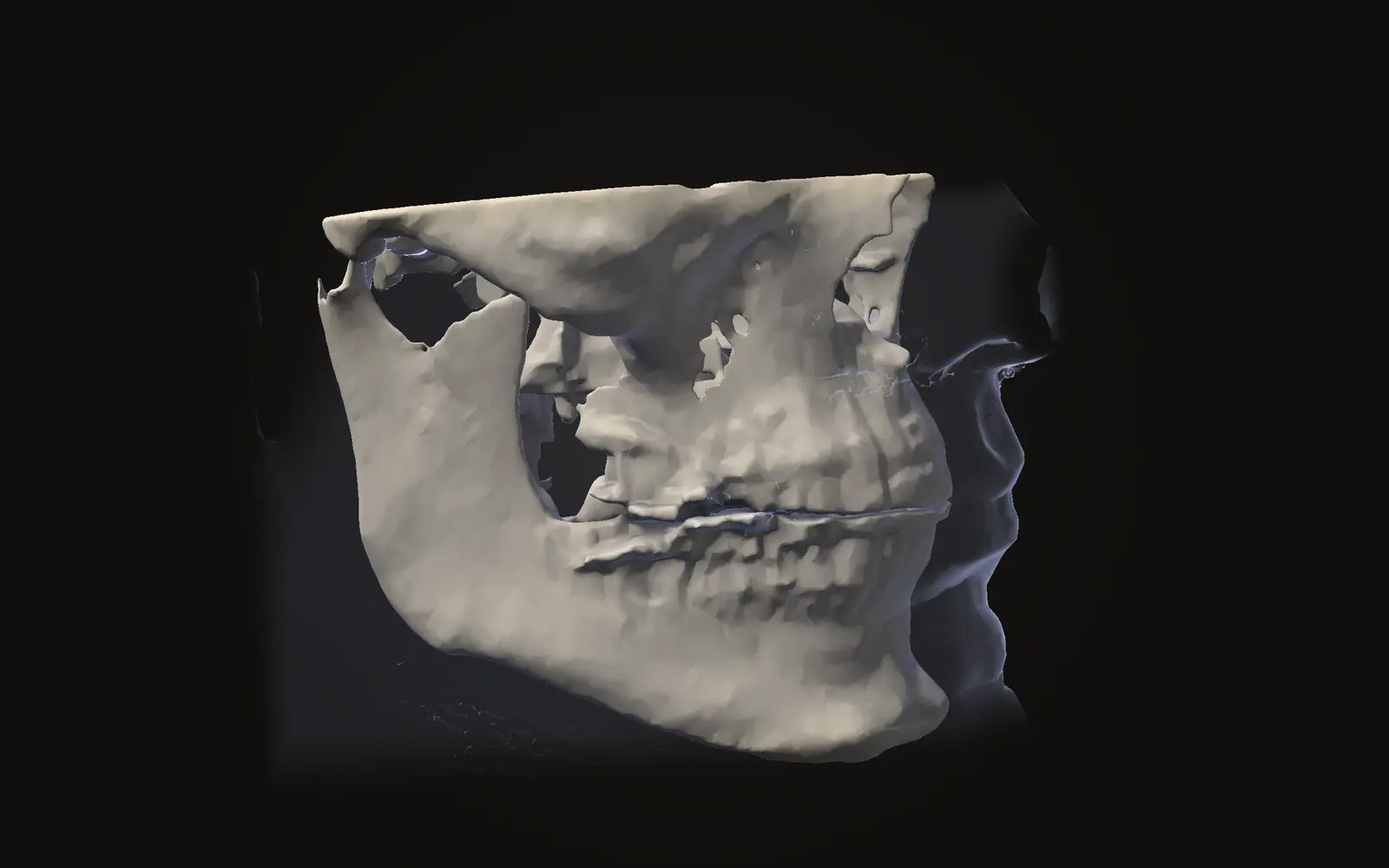
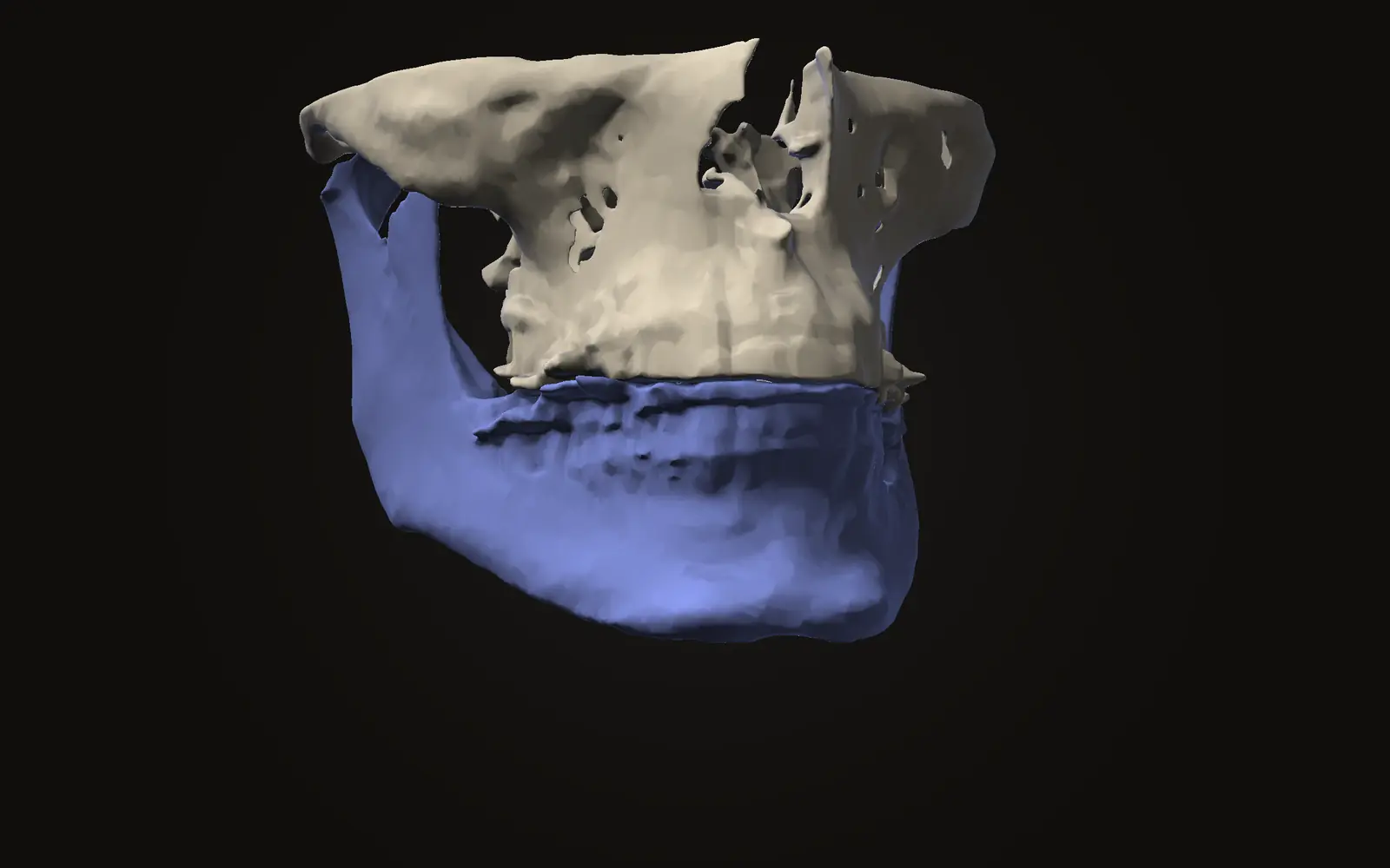
After the line-up, the two surfaces can be coloured by how far apart they are. In the 10 patients scanned before and 1 week after upper jaw surgery, this colour-coded display showed changes in the position of the lower jaw's rami.[1] The average displacement over all surfaces was 0.77 mm, at the condyle 0.70 mm, and the authors noted these values were close to the image resolution of 0.60 mm.[1]
On a coloured map, a displacement of a few tenths of a millimetre sits near the size of a voxel, so the colour alone does not separate a real shift from the limits of the images. Voxel size is explained in CBCT voxel size and sharpness.
Another study checked the fit of the cranial base line-up by eye, in semi-transparent axial, sagittal and coronal slices, before it looked at surface distances. It reported a superimposition error under 0.5 mm in 8 patients with a before and after pair, 4 growing and 4 not growing.[5] Checking in the slices as well as in 3D is a habit worth borrowing for any comparison.
Doing it in Jaw Viewer#
Jaw Viewer opens two scan dates side by side in the browser. You choose the skull base as the reference when both scans include it, limit the region to anatomy the two scans share, and run the automatic alignment. If the bones in the reference region do not agree, you can match point pairs by hand or nudge the alignment yourself. The alignment only turns and shifts one scan; it never bends the images to agree. The steps are in the compare guide.
| View | What it shows |
|---|---|
| Side by side | Each scan in its own view, with cameras and slices kept in step |
| Blend | One scan laid over the other, with a slider for which date shows |
| Swipe | A divider you drag across both scans |
| Checkerboard | Alternating squares from each date |
| Flicker | The view switching between the two dates |
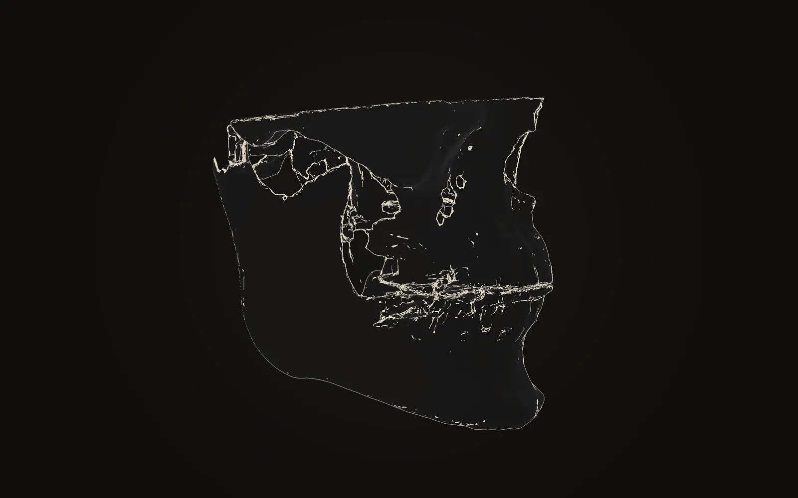
| Difference | Where the two scans differ |
| Distance map | A 3D surface coloured by distance from the other scan's surface |
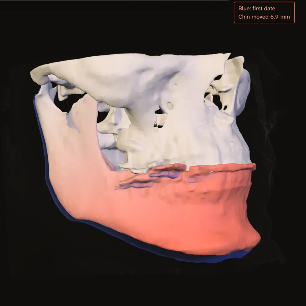
Jaw Viewer does not report how far each jaw piece moved, and it gives no accuracy figure for its alignment. The limits page says what the match depends on. Two scans from different machines, with the head in a different position, or with metal near the jaws, are harder to line up, and a mismatch in the reference region makes unchanged bone look as if it moved.
Did the line-up work?#
Before reading the colours, check the cranial base in several slices and in a blend or checkerboard. If the bone there matches, the colours elsewhere mean something. If it does not, adjust before reading anything.
The method also changes how long this takes. Besides the 10 to 15 s and 3 hour figures above, the 2022 review found run times from a few seconds up to 40 minutes across methods.[4] Software that is automatic and needs one program was the most efficient in that review.[4] The authors of the voxel-based tutorial add a caution: measuring the result quantitatively remains the main challenge of the technique.[3]
To get both scans into the viewer in the first place, see opening a CBCT scan from a CD.
How are before and after jaw surgery scans compared?
One scan is turned and shifted until the cranial base of both scans overlaps, because the operation does not change that bone. What differs elsewhere, such as the jaws, is then shown as an overlay or a coloured distance map.
Why is the skull base used to line up the scans?
Studies of surgical patients use it because the reference area is not affected by growth or by the operation. Teeth and jaws are the parts that moved, so they cannot serve as the reference.
How long does lining up two CBCT scans take?
It depends on the method. One study reported 10 to 15 s for a fast voxel-based method. An older open-source route takes about 3 hours, and commercial software a few minutes.
Can I compare my before and after scans myself?
Jaw Viewer can open both scans in your browser, line them up on the skull base and show the difference. It shows where the two scans differ. It does not say what a difference means.
Sources
- Cevidanes LH, Bailey LJ, Tucker GR Jr, et al. (2005). Superimposition of 3D cone-beam CT models of orthognathic surgery patients. Dentomaxillofac Radiol, 34(6): 369-375. 10 patients, upper jaw surgery only, scans before and 1 week after.DOIPubMed
- Bazina M, Cevidanes L, Ruellas A, et al. (2018). Precision and reliability of Dolphin 3-dimensional voxel-based superimposition. Am J Orthod Dentofacial Orthop, 153(4): 599-606. 31 surgical orthodontic patients with a scan before and after surgery within 12 months.DOIPubMed
- Dot G, Rafflenbeul F, Salmon B (2020). Voxel-based superimposition of Cone Beam CT scans for orthodontic and craniofacial follow-up: Overview and clinical implementation. Int Orthod, 18(4): 739-748. Tutorial and overview, with three illustrative cases.DOIPubMed
- Andriola FO, Haas Junior OL, Guijarro-Martínez R, et al. (2022). Computed tomography imaging superimposition protocols to assess outcomes in orthognathic surgery: a systematic review with comprehensive recommendations. Dentomaxillofac Radiol, 51(3): 20210340. 13 studies, 10 of them using CBCT.DOIPubMed
- Weissheimer A, Menezes LM, Koerich L, et al. (2015). Fast three-dimensional superimposition of cone beam computed tomography for orthopaedics and orthognathic surgery evaluation. Int J Oral Maxillofac Surg, 44(9): 1188-1196. 18 patients, of whom 8 had a pre and post treatment pair.DOIPubMed
Terms used
- Cranial base
- The underside of the skull behind and above the eyes and nose. It is not changed by jaw surgery, so it is the usual reference for lining up before and after scans.
- Superimposition
- Lining up two scans of the same person on a shared reference so that differences elsewhere can be compared. Also called registration.
- Registration
- Lining up two scans of the same person so the same anatomy sits in the same place, before you compare them. The match is judged on a reference region both scans share.
- CBCT
- Cone beam computed tomography. A scanner that rotates once around the head and records a cone-shaped X-ray beam, common in dentistry and jaw surgery.
- Voxel
- A three-dimensional pixel. A scan is a grid of voxels, each with one grey value.
This is general information, not medical advice. Jaw Viewer is not a medical device. Talk to your surgeon, orthodontist or radiologist about your own case.