Key points
- A 2007 review reports that clinically relevant changes of more than 2 mm occur in a surprisingly large percentage of jaw surgery patients from 1 to 5 years after treatment.
- A 2022 systematic review of 132 Class II patients in 4 cohort studies found mandibular advancement and bimaxillary operations highly stable, with low-quality evidence.
- The same review found single-jaw upper jaw operations mostly stable front to back, with problematic stability in the vertical direction.
- A 2016 review of 17 articles linked advancement over 10 mm, counterclockwise rotation and a backward condyle position with a higher risk of condylar resorption.
Relapse is movement of the jaws or teeth back toward their position before the operation. Reviews report that it is not rare: changes of more than 2 mm were found in a surprisingly large percentage of patients between 1 and 5 years after treatment, once the bone had healed.[1] How much moves depends on which jaw was moved, in which direction and by how far.
How is relapse measured?#
Studies compare records taken at set times. The studies in one 2023 review used cephalometric X-ray images and digital models of the teeth.[2] The movement is given in millimetres for the skeleton and the teeth. A 2007 review used 2 mm as its limit for a clinically relevant change.[1]
That limit belongs to those authors. A change under 2 mm is still a change, and a change over 2 mm is not automatically a poor result. The number tells you how reviewers sorted the studies, and nothing about any one person.
Which movements hold best?#
Proffit and colleagues ranked surgical movements in a hierarchy of stability, for operations held with rigid fixation (plates and screws). Moving the chin was very stable, and correcting an asymmetry was stable.[1] Their review adds that fewer patients show long-term change in how the teeth meet than in the bone, because the teeth usually adapt to the skeletal change.[1] The chin operation has its own article.
The same review compared two groups of patients. In the first year, movements for Class II and long-face problems tended to be more stable than those for Class III. From 1 to 5 years, some patients in both groups showed skeletal change, and the Class III group was then the more stable one.[1] Class II means the lower jaw sits behind the upper one. Class III means the opposite.
What does a systematic review of Class II add?#
A 2022 systematic review set out to check the hierarchy for Class II patients. Its authors note that the hierarchy had no systematically reviewed evidence behind it.[3] It found four non-randomised cohort studies with 132 patients in total.[3]
| Operation in Class II | What the review reports |
|---|---|
| Bimaxillary (both jaws) | Highly stable |
| Lower jaw advancement | Highly stable |
| Upper jaw alone | Mostly stable front to back, problematic in the vertical direction |
The authors rate the evidence as low quality and limited, and say more studies are needed.[3] They conclude that it partly supports the hierarchy.[3] The movements are described in the lower jaw operation and the upper jaw operation.
Does relapse happen in most cases?#
A 2023 systematic review of 14 studies concludes that relapse, judged on cephalometric images and digital models, occurs in most cases.[2] That is a broader statement than the Class II review makes. This review included only open-access human studies, most of the studies were retrospective, and the samples were small.[2] Its abstract gives no size of movement. It supports the finding that some movement back is common, but gives no figure for how far.
What is condylar resorption?#
The condyle is the rounded top of the lower jaw where it meets the skull. In condylar resorption it shrinks in shape and volume, and a 2016 systematic review calls it a known factor of surgical relapse.[4] The review included 17 articles. It reports that the condition mainly occurred in women aged 14 to 50 with pre-existing jaw joint dysfunction.[4]
It also reports that three things were associated with a higher risk: advancing the lower jaw by more than 10 mm, rotating it counterclockwise, and repositioning the condyle backward.[4] These associations show which factors appeared together; they do not establish cause and effect.
Why do the numbers differ between reviews?#
The reviews above ask different questions. One ranks movements, one looks only at Class II, one counts relapse in general, and one looks at a single joint problem. Their measures differ as well: skeletal change in millimetres, change in how the teeth meet, or change in the condyle.
The Class II review searched for randomised and controlled trials and included four non-randomised cohort studies.[3] The 2023 review calls most of its studies retrospective with small samples.[2] Treat the figures as the state of the literature, which is still thin.
What can two scans show?#
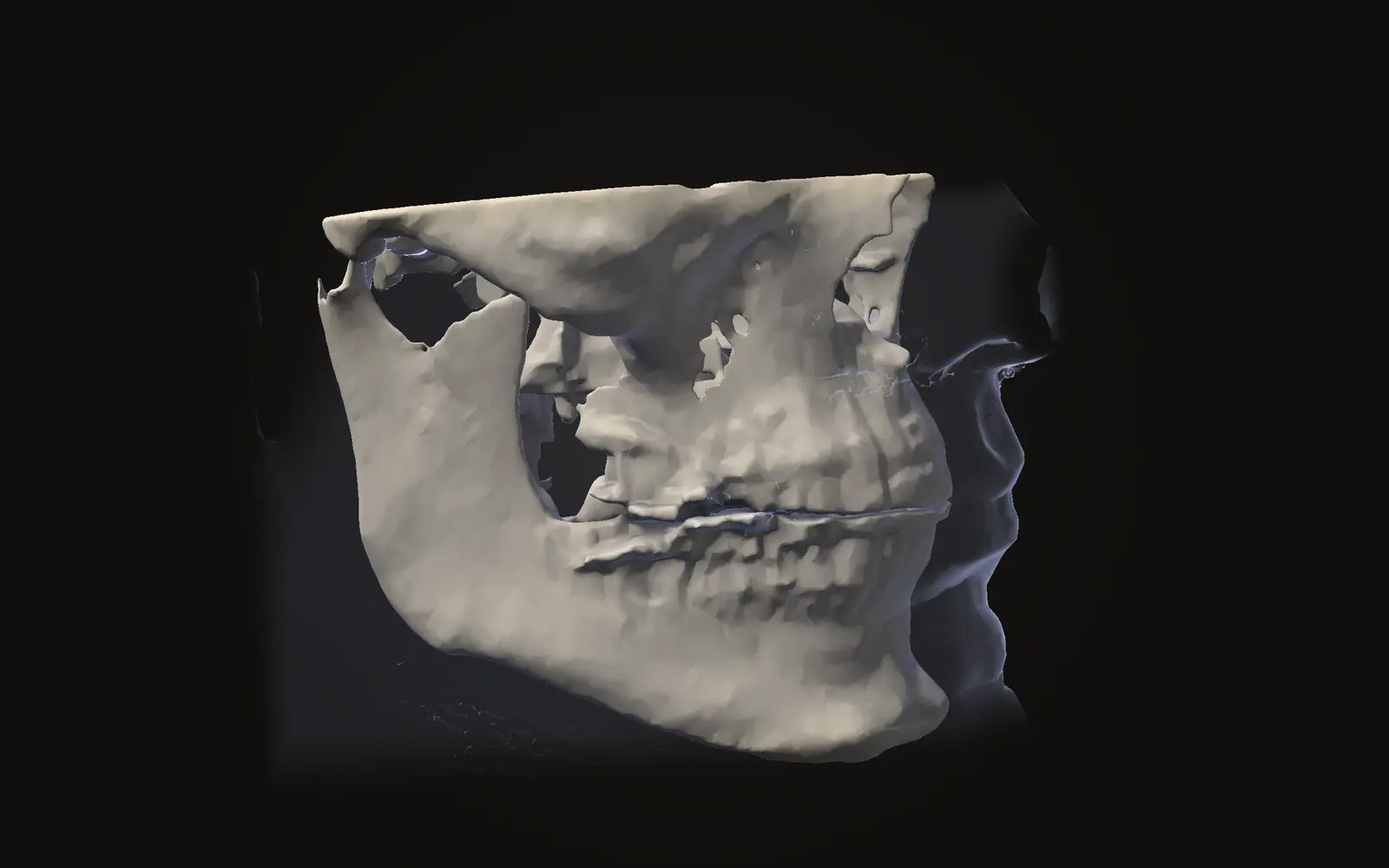
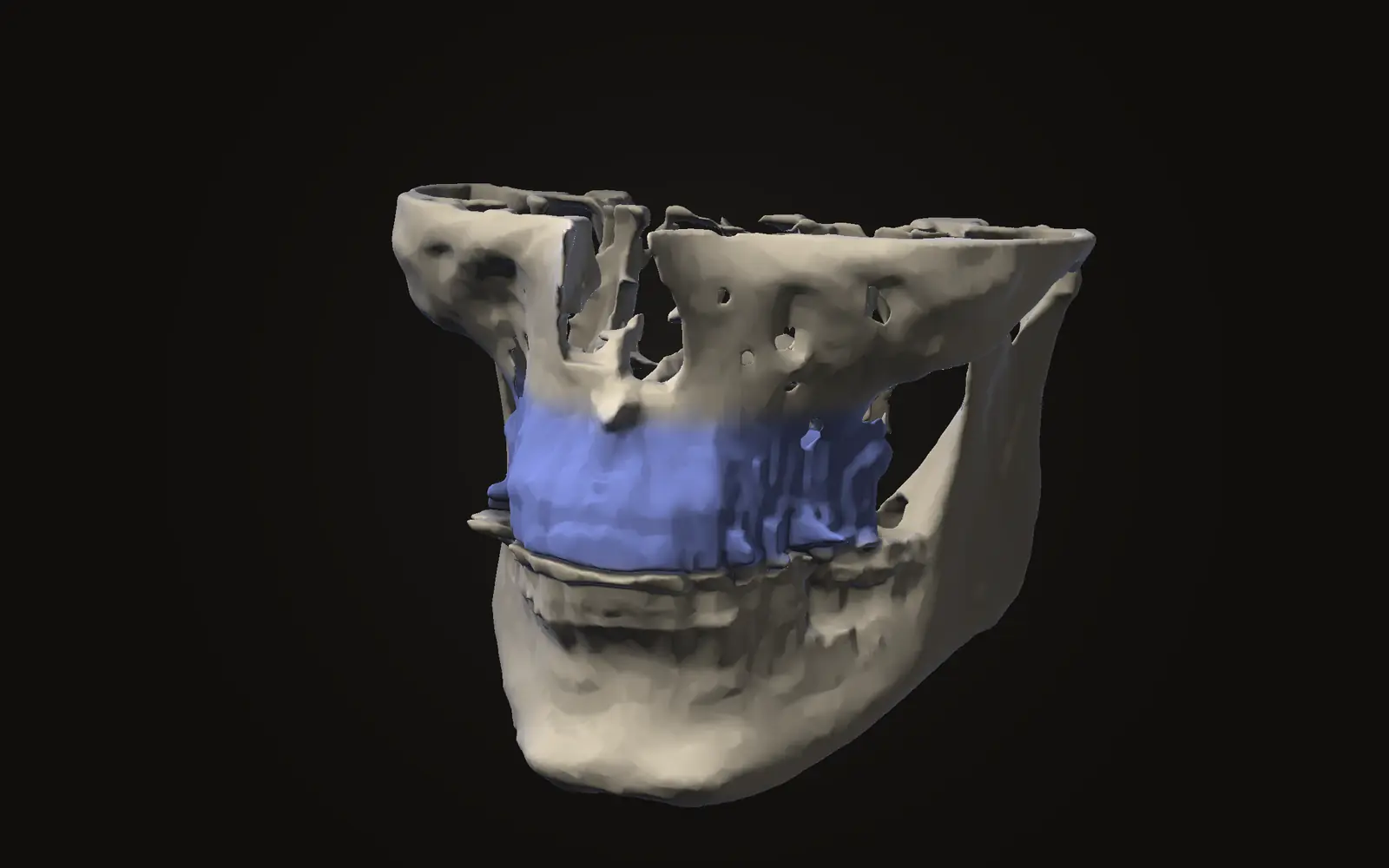
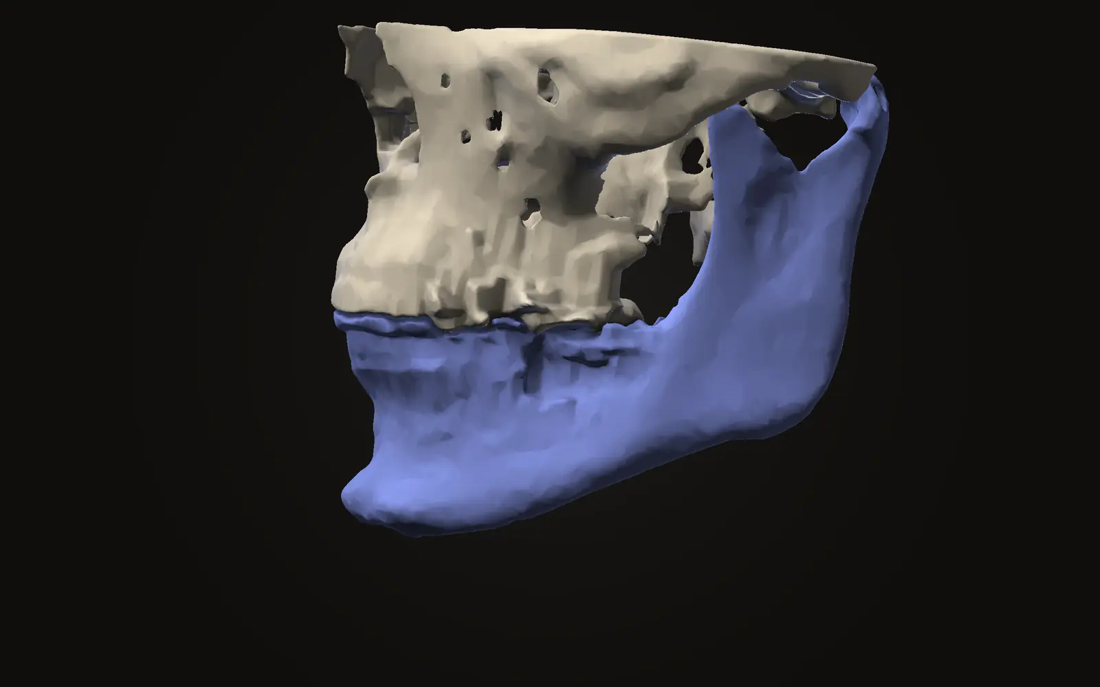
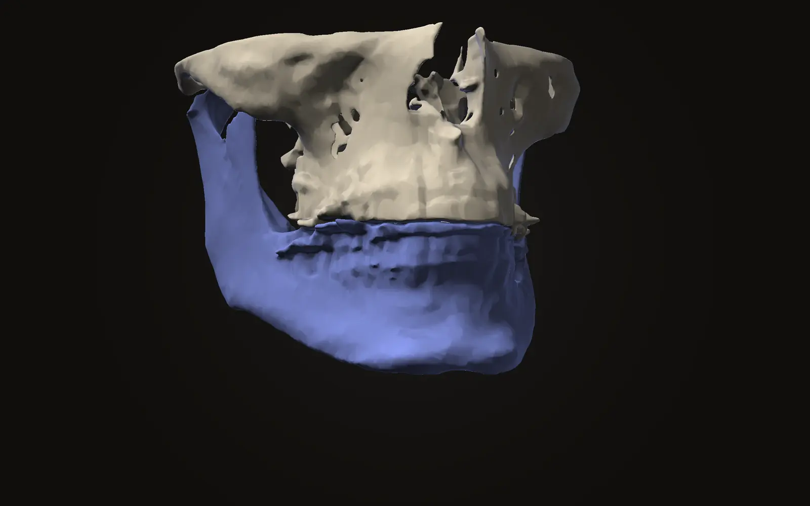
Two scans of the same person, lined up on the skull base, show where the jaws sit on each date. Jaw Viewer can line them up automatically, then show the two dates side by side, blended, swiped, or as a distance map across the bone. Comparing before and after jaw surgery scans explains how the lining up works, and the registration error article explains what limits it.
The viewer shows that a change exists and how big it is, within the limits of both scans. It cannot tell you whether a change is relapse, normal healing or an acceptable result, and it does not tell you what to expect. The guide to comparing scans lists the views.
What is relapse after orthognathic surgery?
Relapse is movement of the jaws or teeth back toward where they were before the operation, after healing. Studies measure it on X-ray images or scans taken at set times after surgery.
How much do the jaws move back after surgery?
It depends on the type of movement. A 2007 review reports changes of more than 2 mm in a surprisingly large percentage of patients between 1 and 5 years. It gives no single figure for everyone.
Is relapse the same as condylar resorption?
No. Condylar resorption is shrinking of the condyle, the top of the lower jaw. A 2016 review calls it a known factor of relapse. Relapse can also have other causes.
Can two scans show whether my jaw moved back?
Two scans lined up on the skull base can show where the jaw sits on each date. Whether a change counts as relapse or a stable result is a clinical judgement the viewer cannot make.
Sources
- Proffit WR, Turvey TA, Phillips C (2007). The hierarchy of stability and predictability in orthognathic surgery with rigid fixation: an update and extension. Head Face Med, 3: 21. Review that ranks surgical movements by stabilityDOIPubMed
- Inchingolo AM, Patano A, Piras F, et al. (2023). Orthognathic surgery and relapse: a systematic review. Bioengineering (Basel), 10: 1071. 14 studies; included only open-access human studiesDOIPubMed
- Eckmüller S, Paddenberg E, Hiller KA, et al. (2022). Relapse in class II orthognathic surgery: a systematic review. BMC Oral Health, 22: 605. 4 non-randomised cohort studies, 132 patients; low quality of evidenceDOIPubMed
- Catherine Z, Breton P, Bouletreau P (2016). Condylar resorption after orthognathic surgery: a systematic review. Rev Stomatol Chir Maxillofac Chir Orale, 117: 3-10. 17 included articlesDOIPubMed
This is general information, not medical advice. Jaw Viewer is not a medical device. Talk to your surgeon, orthodontist or radiologist about your own case.