Key points
- A Le Fort I osteotomy cuts the upper jaw free from the nasal wall back to the pterygomaxillary junction, so it can be moved in all three planes.
- A review of 996 maxillary osteotomies reported neurosensory disturbance in 64.7% of operations, with the evidence rated low. The figure pools studies that define it differently.
- A 2026 review of ischaemic complications found rates of 0.2% to 3.3% in cohort studies, and calls loss of bone blood supply exceptionally rare.
- Reviews of movements and fixation report that changes over 2 mm occur in a surprisingly large share of patients one to five years after surgery.
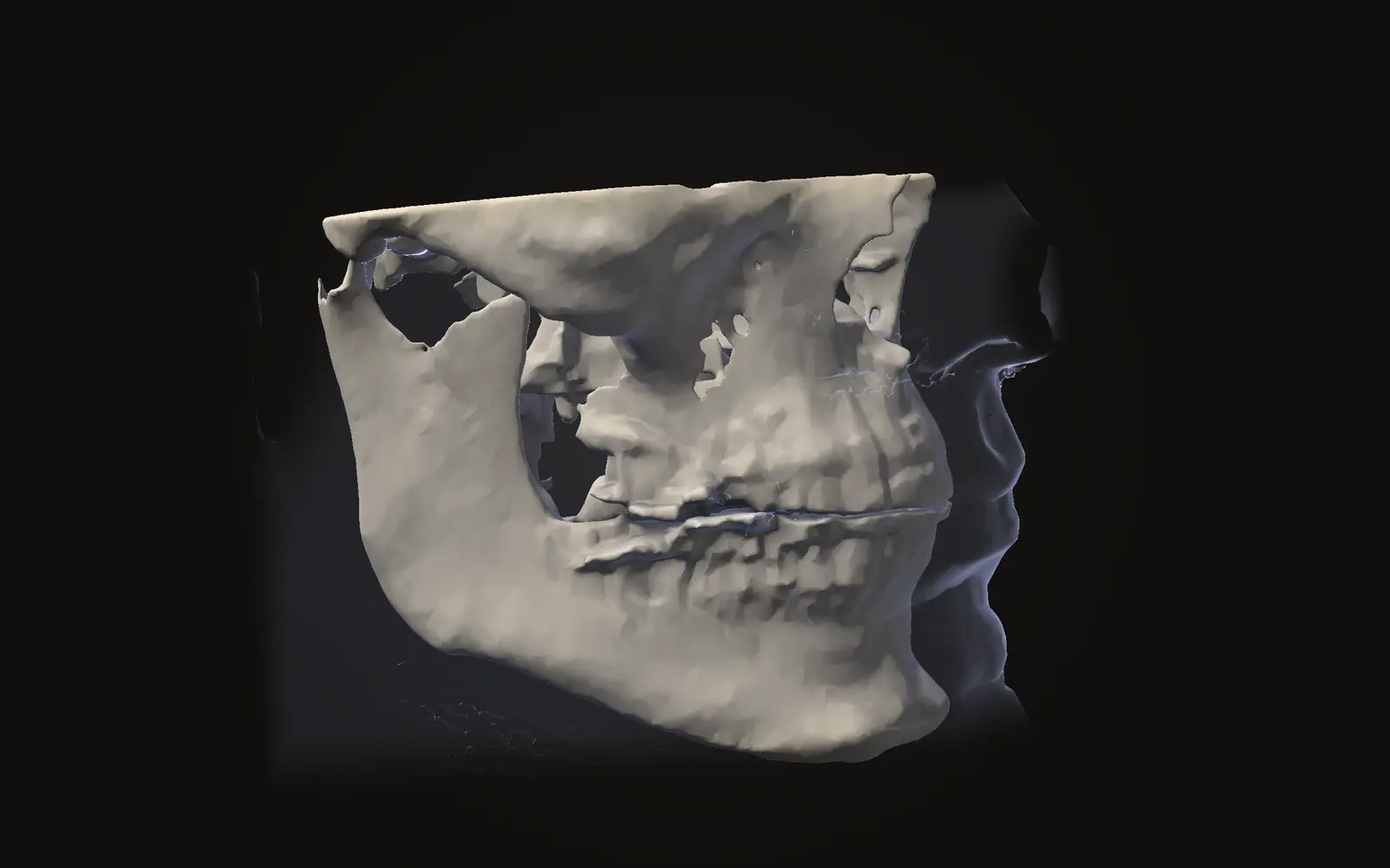
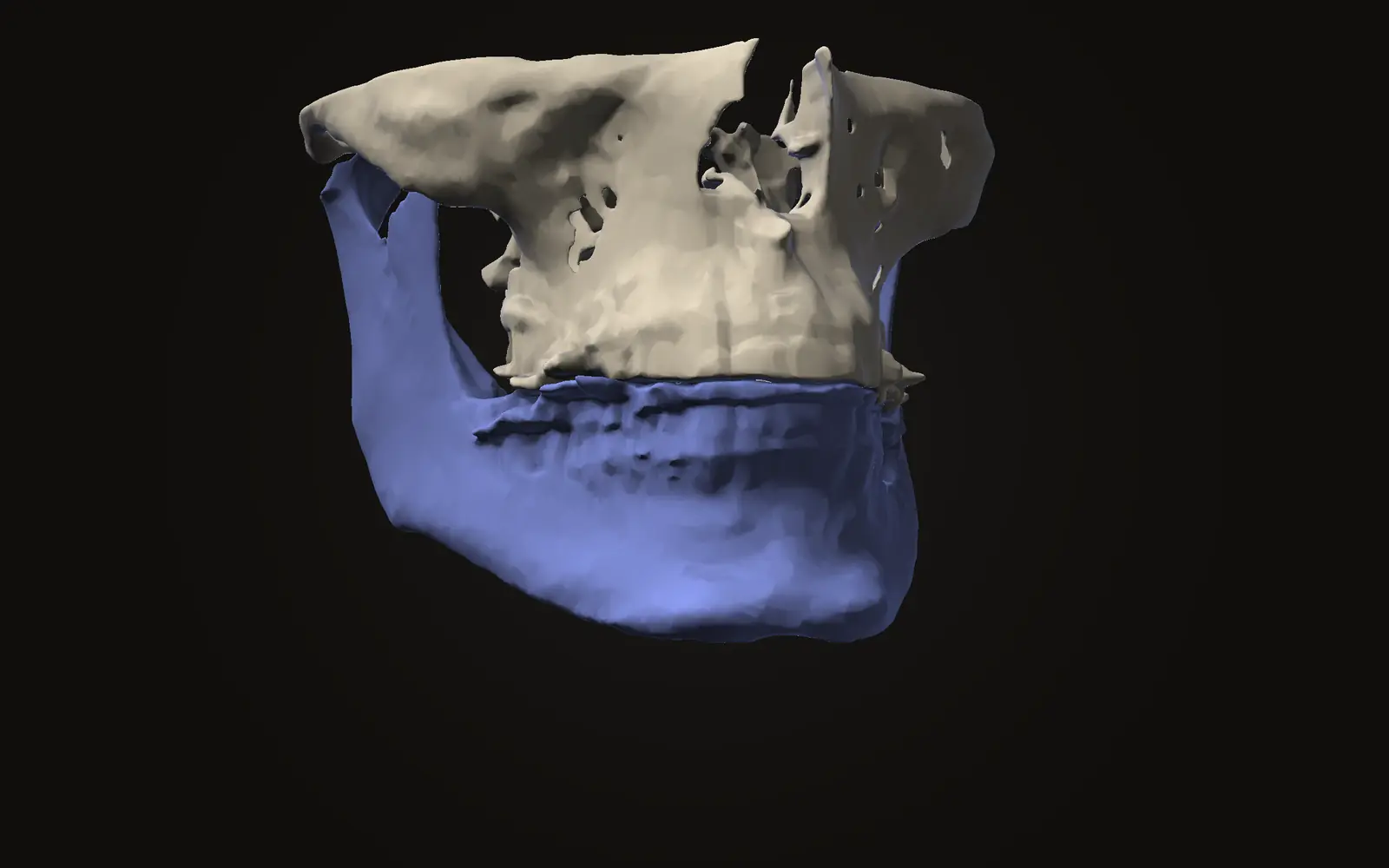
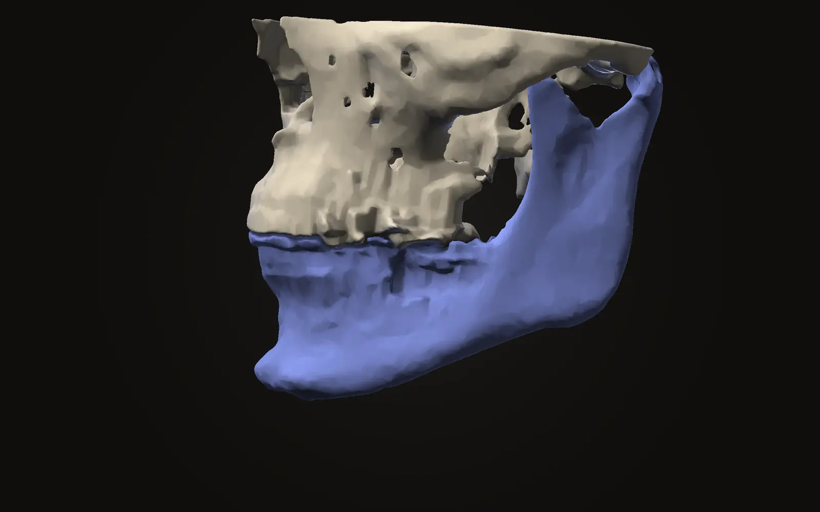
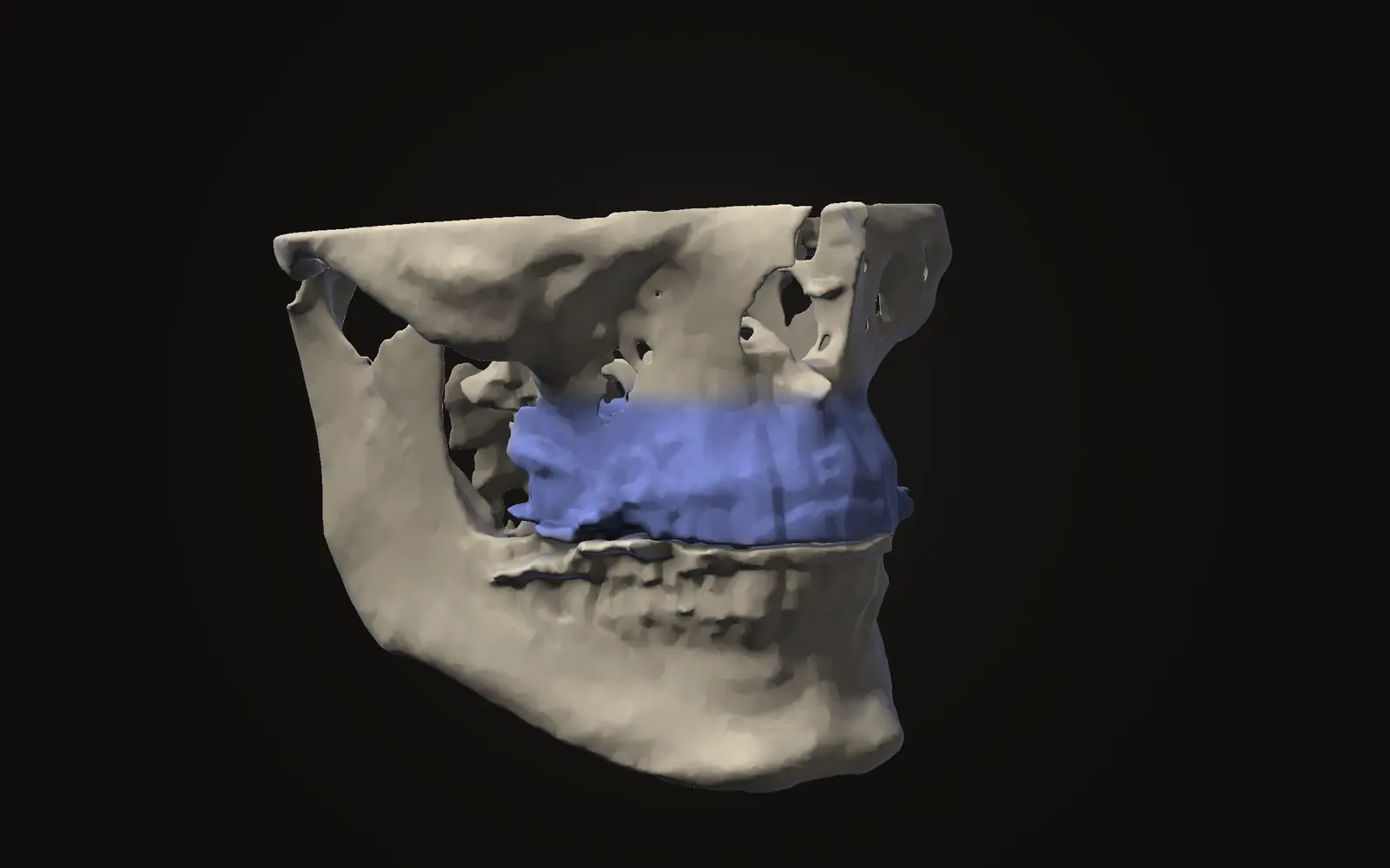
A Le Fort I osteotomy is an operation that cuts the upper jaw free so the part that carries the teeth can be moved and fixed in a new position. The standard cut runs from the lateral wall of the nasal cavity back to the pterygomaxillary junction, and it lets the jaw move in all three planes.[1] Reviews report on stability and complications, though the evidence has limits.
What the operation does#
The upper jaw is the maxilla. In a Le Fort I osteotomy the surgeon separates it from the middle of the face. The standard version also breaks the nasal septum, which frees the tooth-bearing part of the jaw to move in all three planes.[1]
The reviewed literature names three reasons for the operation: an underdeveloped middle third of the face, a maxilla that is too tall, and facial asymmetry.[1] It is one osteotomy among several. Reviews of orthognathic treatment describe it as a team effort between orthodontics and oral and maxillofacial surgery, and note that the timing of treatment and the size of the movements affect how stable the result is.[2] The overview in what orthognathic surgery is places the upper jaw operation next to the lower jaw and chin operations.
A segmental version cuts the upper jaw into more than one piece. It has its own stability pattern, covered below.[3]
Where the name comes from#
The Le Fort I fracture pattern was first described by René Le Fort in 1901. Wasmund first used it in the context of facial orthopaedics in 1921.[1] The operation keeps the name of the fracture.
What do reviews report about complications?#
One review pooled 996 maxillary osteotomies, 864 with conventional cutting tools and 132 with a piezoelectric device. It recorded 156 complication events.[4]
| Complication | Reported prevalence |
|---|---|
| Neurosensory disturbance | 64.7% |
| Haemorrhage | 8.3% |
| Oroantral communication | 7.7% |
| Soft tissue injury | 7.7% |
| Tooth injury | 5.1% |
| Infection | 3.2% |
| Osteonecrosis | 1.9% |
| Permanent nerve injury | 1.3% |
The review grades the evidence as low. Its main question was whether a piezoelectric device changes complication rates, which the authors say was hard to compare with the available literature.[4] A neurosensory disturbance is a change in feeling, and the review lists permanent nerve injury separately, at 1.3%.[4] An oroantral communication is an opening between the mouth and the maxillary sinus.
Segmental Le Fort I: what holds and what does not#
A systematic review of 23 studies looked at the split version. It found that segmental Le Fort I gives stable results in the sagittal plane, which is the front-to-back direction. In the transverse plane, the side-to-side direction, it is less stable for the teeth than for the bone. After a downward movement, the back part of the jaw has little stability.[3]
On complications, the same review found oral fistula and damage to neighbouring teeth were the most frequently described, and postoperative infection the most prevalent at 32.62%.[3] The review also states that all studies on complications had a high potential risk of bias, and the infection figure should be read with that in mind.[3]
How rare is loss of blood supply to the jaw?#
Maxillary avascular necrosis is the complication people fear most, and the 2026 systematic review of seven studies calls it exceptionally rare but serious. Among cohort studies, clinically significant ischaemic complications were reported at 0.2% to 3.3%.[5] The review found no single factor that explained every case, and the link with injury to one artery was inconsistent.[5] A separate scoping review of 33 studies lists the vascular problems reported after the operation, with pseudoaneurysm the most common.[1]
Does the new position hold?#
Orthognathic movements differ in how stable they are, and Proffit and colleagues ranked them in a hierarchy. Their review also states that changes of more than 2 mm occur in a surprisingly large percentage of patients from one to five years after treatment, once surgical healing is complete.[6] In the first year after surgery, movements for Class II and long-face problems tended to be more stable than those for Class III.[6] Relapse after jaw surgery goes through that evidence in more detail.
What changes in the face#
The upper jaw sits under the nose and the upper lip, so the soft tissue there can change. Two companion articles cover it: how lips and chin follow bone, and what studies measured about nose width. The lower jaw operation has its own article, the sagittal split osteotomy.
What a scan shows of the upper jaw#
3D imaging in orthognathic surgery covers how scans are used around jaw operations. On your own scan, the upper jaw, the roots of the teeth and the maxillary sinuses are visible in slices and in 3D. Jaw Viewer can outline both sinuses and give their volumes, as the airway and sinuses guide explains. It does not show how an operation would go, and it cannot estimate a risk.
What is a Le Fort I osteotomy?
An operation that cuts the upper jaw free so the part carrying the teeth can be moved and fixed in a new position. It is one of the standard upper jaw operations in orthognathic treatment.
Is a Le Fort I osteotomy the same as a Le Fort fracture?
The name comes from a facial fracture pattern described by René Le Fort in 1901. The operation was first used for jaw correction by Wasmund in 1921, according to a 2025 review.
How common is numbness after a Le Fort I osteotomy?
One review pooled 996 maxillary osteotomies and reported neurosensory disturbance in 64.7%. Studies define it differently and the evidence is rated low, so the figure describes a pool of studies, not one person's chance.
Can I see my upper jaw and sinuses on my own CBCT scan?
Yes. Slices in three directions and a 3D view show the upper jaw, teeth and both maxillary sinuses. The viewer can outline the sinuses and give their volume. It cannot judge an operation or a risk.
Sources
- Alves LC, Alves JM, Soares AM, et al. (2025). Vascular complications related to Le Fort I osteotomy: a scoping review. Med Oral Patol Oral Cir Bucal, 30: e830-8. Scoping review of 33 studiesDOIPubMed
- Malik OH, Waring DT, Lloyd R, et al. (2016). An overview of the surgical correction of dentofacial deformity. Dent Update, 43: 550-62. Evidence-based overview written for general dentistsDOIPubMed
- Haas Junior OL, Guijarro-Martínez R, de Sousa Gil AP, et al. (2017). Stability and surgical complications in segmental Le Fort I osteotomy: a systematic review. Int J Oral Maxillofac Surg, 46: 1071-87. 23 studies; all complication studies at high potential risk of biasDOIPubMed
- Thereza-Bussolaro C, Galván Galván J, Pachêco-Pereira C, et al. (2019). Maxillary osteotomy complications in piezoelectric surgery compared to conventional surgical techniques, a systematic review. Int J Oral Maxillofac Surg, 48: 720-31. 996 maxillary osteotomies; low level of evidenceDOIPubMed
- Lee CW, Hwang SY, Esmaeili L, et al. (2026). Maxillary avascular necrosis after Le Fort I osteotomy: a systematic review of ischemic complications. Maxillofac Plast Reconstr Surg, 48: 23. Seven studiesDOIPubMed
- Proffit WR, Turvey TA, Phillips C (2007). The hierarchy of stability and predictability in orthognathic surgery with rigid fixation: an update and extension. Head Face Med, 3: 21.DOIPubMed
Terms used
- Maxilla
- The upper jaw bone. It carries the upper teeth and forms the roof of the mouth, the floor of the nose and part of the cheekbone area.
- Le Fort I osteotomy
- A planned cut through the upper jaw above the teeth, so the whole tooth-bearing part can be moved and fixed in a new position.
- Osteotomy
- A planned cut in a bone.
- Neurosensory disturbance
- A change in feeling, such as numbness or tingling, in the area a nerve supplies.
- Oroantral communication
- An opening between the mouth and the maxillary sinus.
- Avascular necrosis
- Death of bone or tooth tissue because its blood supply is lost.
This is general information, not medical advice. Jaw Viewer is not a medical device. Talk to your surgeon, orthodontist or radiologist about your own case.