Key points
- A 2026 meta-analysis of 47 studies (5,406 patients) pooled 21% (95% CI 13% to 32%) with some sensation change at 12 months after sagittal split osteotomy.
- A 2007 review of 7 studies found impairment in 63.3% of patients at day 7 and 12.8% at 1 year by objective tests; patient reports gave 83% and 18.4%.
- A 2016 review of 59 studies pooled a mean of 33.9% per patient for lower-lip sensation changes, counted across studies that each defined and timed the measurement differently.
- In a review of 61 reports, 16 (26%) did not state the incidence of nerve injury and 22 (36%) did not assess the nerve before the operation.
- A review of 7 imaging studies found general agreement that less bone between the canal and the outer cheek-side cortex goes with more frequent sensation changes, but no standard way of measuring that distance.
Many people who have a lower jaw operation notice changed feeling in the lower lip and chin afterwards. Reviews of sagittal split osteotomy (BSSO) report this in 12.8% to 33.9% of patients, and the figures differ because the studies measured at different times and in different ways.[1][2] The reviews counted different kinds of sensation change, and evidence on who is affected remains limited.
What is being counted?#
Studies call the change in feeling a neurosensory disturbance. The reviews link it to injury of the inferior alveolar nerve, which runs through the mandibular canal of the lower jaw.[3] The operation itself is described in the BSSO article, and the canal in the nerve canal on CBCT.
A 2016 review of 59 studies lists lower-lip neurosensory disturbance among the four most common complications of BSSO, with a pooled mean of 33.9% per patient.[2] Other reviews give lower figures because they pool different measures and time points.
Which figure is the right one?#
Each figure answers a slightly different question.
| Review | What it counted | Figure |
|---|---|---|
| Colella 2007, 7 studies | Impairment by objective tests, 1 year | 12.8%[1] |
| Colella 2007 | Impairment by patient report, 1 year | 18.4%[1] |
| Bertagna 2026, 47 studies, 5,406 patients | Any disturbance reported or assessed at 12 months | 21% (95% CI 13% to 32%)[3] |
| Verweij 2016, 59 studies | Pooled mean, per patient, across study time points | 33.9%[2] |
The 2026 meta-analysis defined persistent disturbance as any disturbance reported or assessed at 12 months.[3] Its 95% confidence interval runs from 13% to 32%.[3]
How does timing change the picture?#
The 2007 review adds the time dimension. At day 7, impairment by objective tests was found in 63.3% of patients, and by patient report in 83%. At 1 year the figures were 12.8% and 18.4%.[1] Sensation changes are common in the first week and far less common a year later in that review.
The same review compared the two methods. Patient reports gave a higher frequency than objective tests at every follow-up time it considered.[1] Agreement between them, measured with Cohen's kappa, was 0.53 at 1 week and 0.90 at 1 year.[1] The authors conclude that objective tests are the most sensitive within 3 months of the operation.[1]
Neither review says how any one person's sensation will change over time.
Why is it so hard to compare studies?#
A review of 61 reports published between 1990 and 2013 looked at how the studies assessed the nerve. In 16 reports (26%) the incidence of injury was not stated. In 22 (36%) the nerve was not assessed before the operation.[4] Subjective evaluation was the most common method.[4] The authors conclude that the wide variation in reported incidence is due to a lack of standardised assessment and reporting, and call for an international consensus on the method.[4]
The 2026 meta-analysis ends on the same point: standardised assessments and adequately powered studies are needed.[3]
What do studies say about risk factors?#
The 2026 meta-analysis looked at factors in the included studies. Older age was significantly associated with persistent disturbance in 5 of 9 studies, greater mandibular advancement in 2 of 2, and exposure or handling of the nerve during the operation in 2 of 3.[3] Sex, skeletal class, fixation method, third molar presence and a genioplasty at the same operation were not significantly associated in most of the studies that examined them (for example, sex in 8 of 9 studies and genioplasty in 3 of 5).[3] Some of these factors rest on very few studies, as the counts show.
What does the position of the canal have to do with it?#
A 2014 review asked whether the position of the mandibular canal on a 3D scan before the operation relates to sensation changes after it. Seven of 69 articles met its criteria.[5] The general consensus was that the less space between the canal and the outer border of the cheek-side cortex, the more frequent the disturbance. Greater bone density also appeared to go with a higher incidence.[5]
The review also found that there was no standard way of measuring canal position, or of evaluating the disturbance. It concludes that more studies are needed to confirm the relationship between the position of the nerve and the outcome.[5]
What the viewer can show#
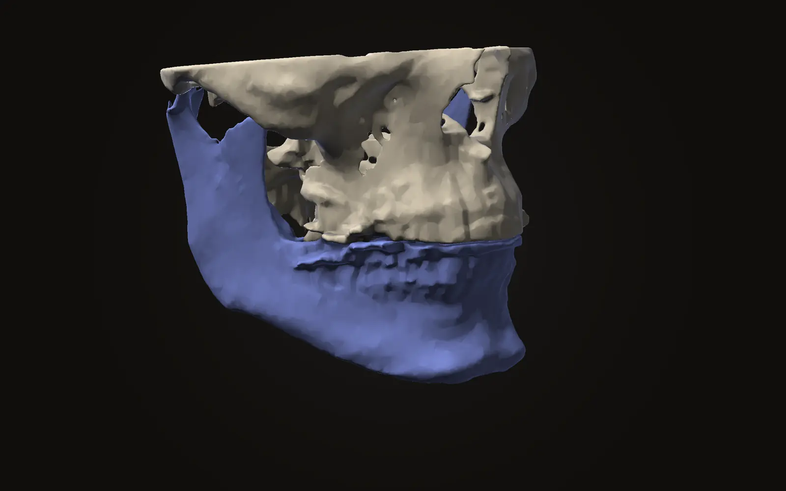
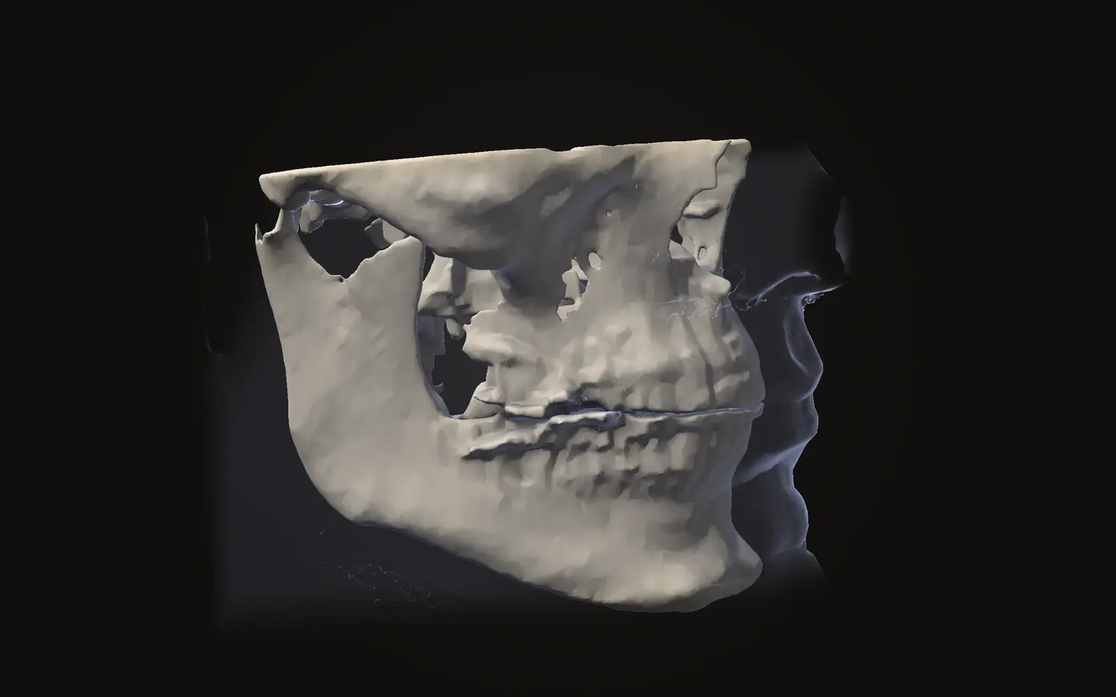
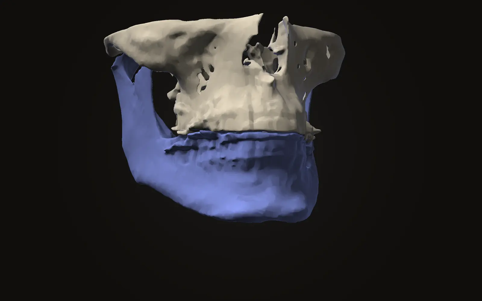
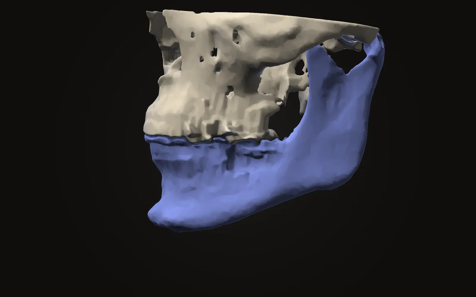
On a CBCT scan, Jaw Viewer lets you scroll the lower jaw in slices in three directions, turn it in 3D, trace the nerve canal by hand and measure distances in millimetres. The guide to viewing and measuring covers the tools, and the limits of how closely the canal can be followed are set out in the nerve canal on CBCT.
The viewer shows where the canal runs in the images. It says nothing about nerve function and nothing about what the figures above mean for your case. Your surgical team can explain that with your history and your scan in front of them.
How common is numbness of the lip after BSSO?
A 2026 meta-analysis of 47 studies pooled 21% of patients with some sensation change at 12 months. Earlier reviews give 12.8% to 33.9%, depending on timing and on how sensation was measured.
Why do studies give such different numbness figures?
They differ in when they measured, whether they used objective tests or patient reports, and how they defined a change. A review of 61 reports links the wide range to non-standard assessment and reporting.
Does a scan show how close the nerve canal is to the bone surface?
Yes, the canal can be followed in CBCT slices. A review of 7 studies found general agreement that a thinner layer of bone outside the canal goes with more sensation changes, but no standard way of measuring it.
Does the viewer tell me how likely numbness is for me?
No. It shows where the canal runs in the scan and lets you measure distances. It says nothing about nerve function or about what the figures in studies mean for one person.
Sources
- Colella G, Cannavale R, Vicidomini A, et al. (2007). Neurosensory disturbance of the inferior alveolar nerve after bilateral sagittal split osteotomy: a systematic review. J Oral Maxillofac Surg, 65: 1707-15. Seven studies.DOIPubMed
- Verweij JP, Houppermans PN, Gooris P, et al. (2016). Risk factors for common complications associated with bilateral sagittal split osteotomy: a literature review and meta-analysis. J Craniomaxillofac Surg, 44: 1170-80. Literature review and meta-analysis of 59 studies.DOIPubMed
- Bertagna AE, Van der Cruyssen F, Miloro M (2026). Persistent neurosensory disturbance following sagittal split osteotomy: a systematic review and meta-analysis of one-year outcomes and risk factors. J Oral Maxillofac Surg, 84: 25-44. 47 studies, 5,406 patients, searched up to February 2025.DOIPubMed
- Agbaje JO, Salem AS, Lambrichts I, et al. (2015). Systematic review of the incidence of inferior alveolar nerve injury in bilateral sagittal split osteotomy and the assessment of neurosensory disturbances. Int J Oral Maxillofac Surg, 44: 447-51. 61 reports published between 1990 and 2013.DOIPubMed
- Rich J, Golden BA, Phillips C (2014). Systematic review of preoperative mandibular canal position as it relates to postoperative neurosensory disturbance following the sagittal split ramus osteotomy. Int J Oral Maxillofac Surg, 43: 1076-81. Seven of 69 articles met the inclusion criteria.DOIPubMed
Terms used
- BSSO
- Bilateral sagittal split osteotomy. A planned cut along both sides of the lower jaw that lets the tooth-bearing part slide forward or back.
- Neurosensory disturbance
- A change in feeling, such as numbness or tingling, in the area a nerve supplies.
- Inferior alveolar nerve
- The nerve that runs through a canal inside the lower jaw and supplies feeling to the lower teeth, lip and chin.
This is general information, not medical advice. Jaw Viewer is not a medical device. Talk to your surgeon, orthodontist or radiologist about your own case.