Key points
- Soft tissue moves by a fraction of the bone movement. One review reports about 0.6 mm of upper lip movement per 1 mm of tooth movement after a Le Fort I advancement without an alar base cinch suture, and 0.9 mm with one.
- People differ widely around the average ratios, so a ratio describes a group, not one person.
- The latest systematic review of 3D studies found no data that can be applied directly in daily practice, so a face prediction is an estimate with a range.
When a surgeon moves a jaw, the skin and lips move too, but by less than the bone and in a pattern that differs from person to person. Researchers describe this with soft-to-hard tissue ratios. This article covers what the systematic reviews found for the three most common moves, and what they could not settle.
How to read a ratio#
A ratio of 0.6:1 means a point on the lip moves 0.6 mm for each 1 mm the matching tooth or bone point moves. Ratios are averages across patients in published studies. They come from measuring soft tissue before and after surgery on the same people, so they describe groups, not one face.
Upper jaw, Le Fort I#
A review of the studies on moving the upper jaw (37 articles in all, only three of them prospective) found a high spread in the ratios that mostly disappeared once studies were grouped by confounding factors. With the available data, it suggested a ratio of 0.6:1 from the tooth tip to the upper lip when the surgeon does not use an alar base cinch suture, and 0.9:1 when one is used.[1] The cinch is a stitch that narrows the base of the nose, and the review ties it to the difference.
So advancing the upper incisors by 4 mm would, on those averages, move the upper lip about 2.4 mm without the stitch and 3.6 mm with it. That is arithmetic on the review's averages, not a prediction for anyone.
Lower jaw, sagittal split#
For lower-jaw advancement, a review of 12 studies (three prospective) found the lower lip moved about half as far as the lower incisors. The skin at the chin (soft tissue pogonion) and the crease under the lip (mentolabial fold) moved about as far as their bone points, a 1:1 ratio, with one trend towards more than 1:1 in the long term. The upper lip mostly moved back, with high variability.[2]
For setback, a review of eight studies (two prospective) found a 1:1 ratio between lower lip and lower incisors in the long term, a little lower in the short term, and a 1:1 ratio for the crease under the lip. The chin skin followed the chin bone 1:1 in the short term, with a trend to less in the long term. The upper lip mostly moved forward, again with large variation.[3]
Both reviews reach the same cautious conclusion: evidence-based conclusions on soft tissue change are hard to draw, because many studies were retrospective, designs were weak, and outcomes were measured in different ways. They ask for prospective studies that leave out patients who had extra surgery such as a chin operation or upper-jaw surgery.[2][3]
Chin, genioplasty#
A review of chin procedures included 33 articles (22 found by search and 11 more in reference lists), three of them prospective. It suggested a ratio of 0.9:1 between chin skin and bone for chin advancement and 0.55:1 for chin setback.[4] As with the upper jaw, the variation across studies largely disappeared when results were grouped by confounding factors, and the authors call for more prospective studies grouped that way.[4]
What three-dimensional studies add#
A 2025 review looked specifically at studies from 2017 to 2023 that measured in three dimensions. From 897 articles it kept 10, all of which compared before and after scans by overlaying them. Results for the area around the nose, the upper lip and the chin were ambiguous, and the authors state that there are no data that can be applied to daily clinical practice. The limited evidence published in 2017 remains.[5]
Where this leaves a prediction#
The reviews give workable averages with a large spread between people. The 3D evidence is thinner than the 2D evidence. A tool that draws a single confident face would claim more than the research supports, so treat any picture of a predicted face as one possible outcome.
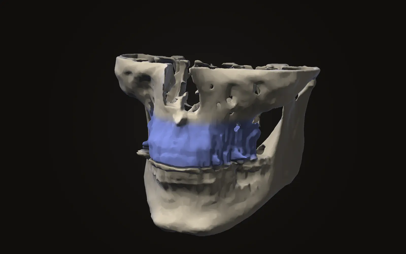
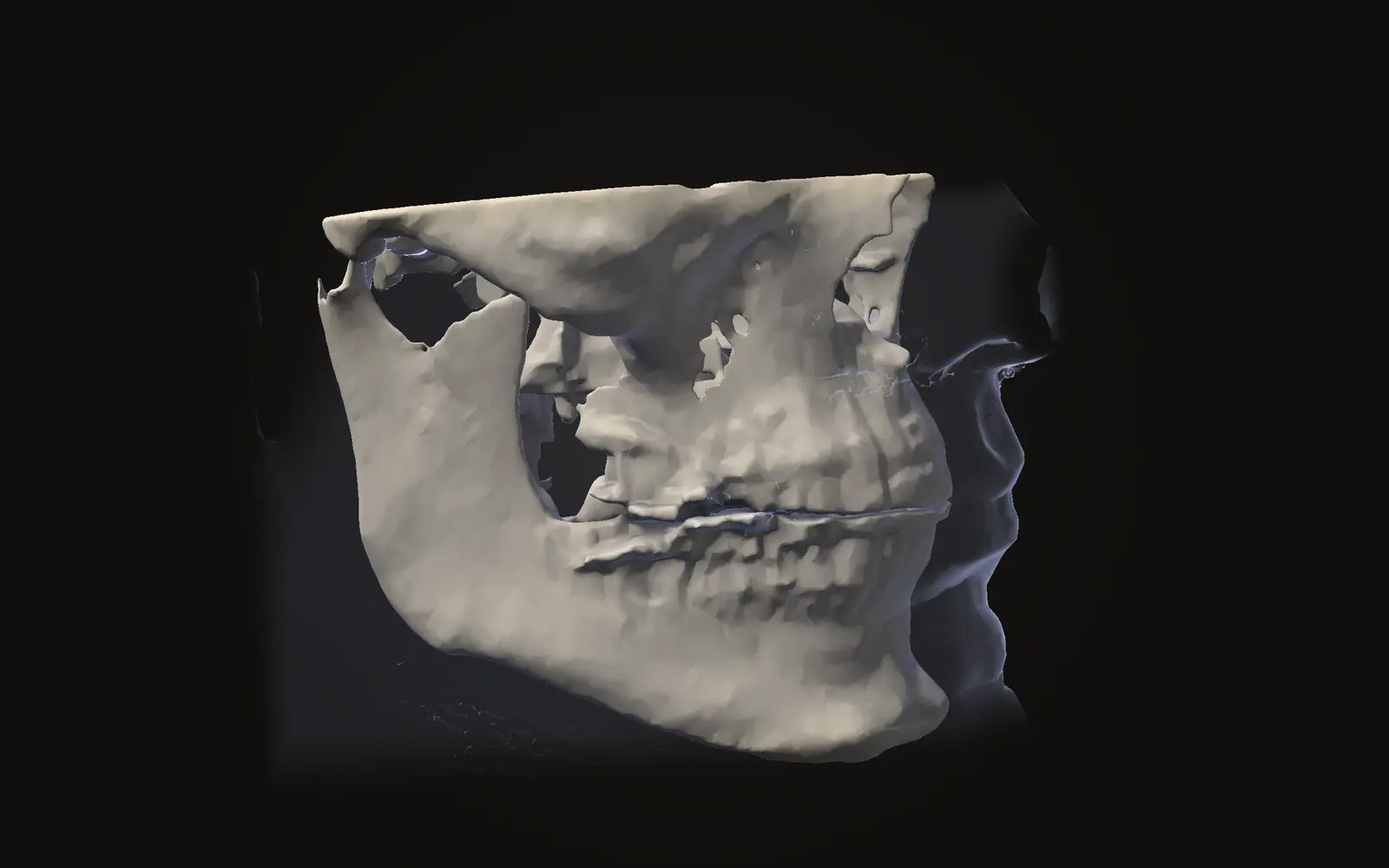
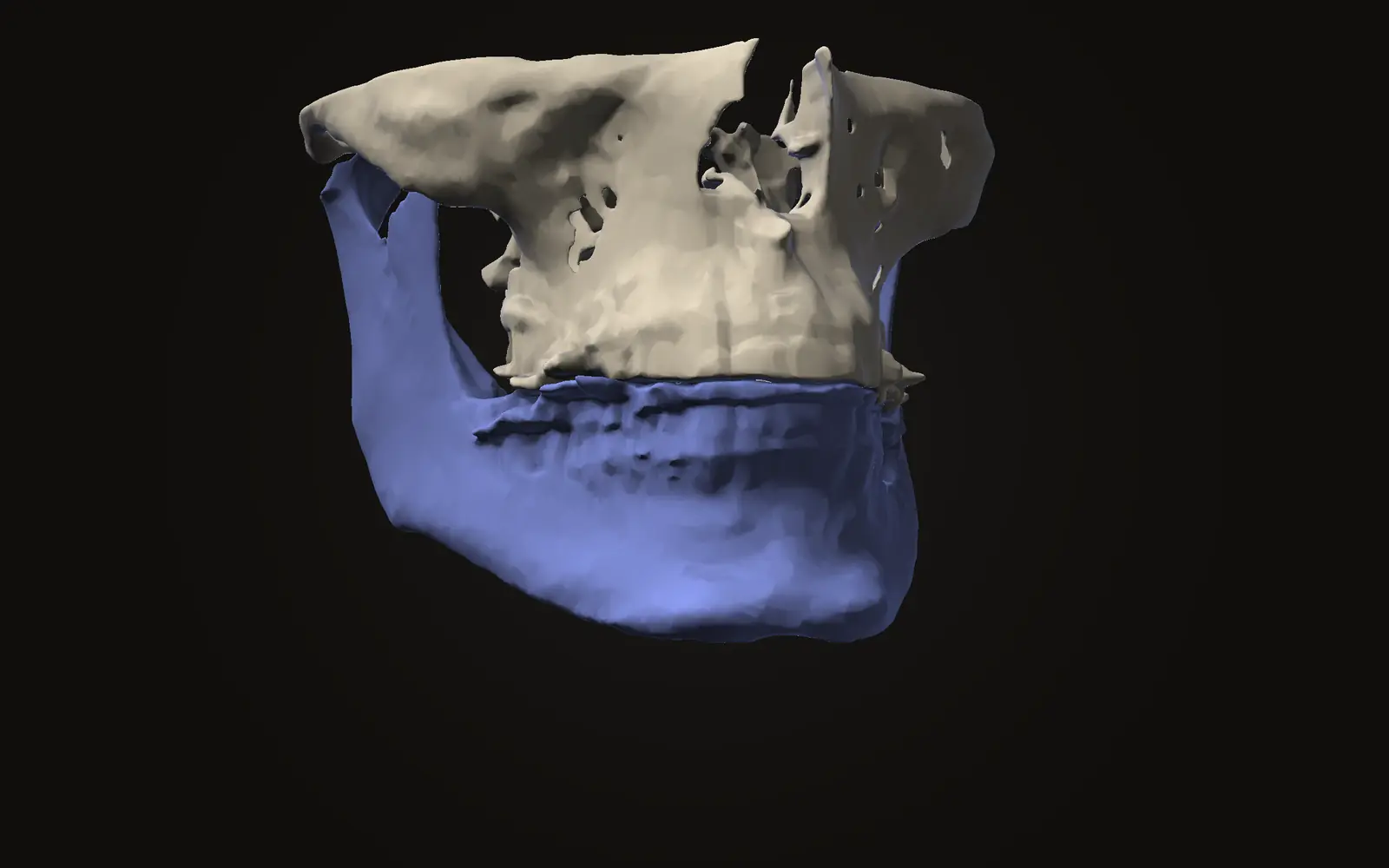
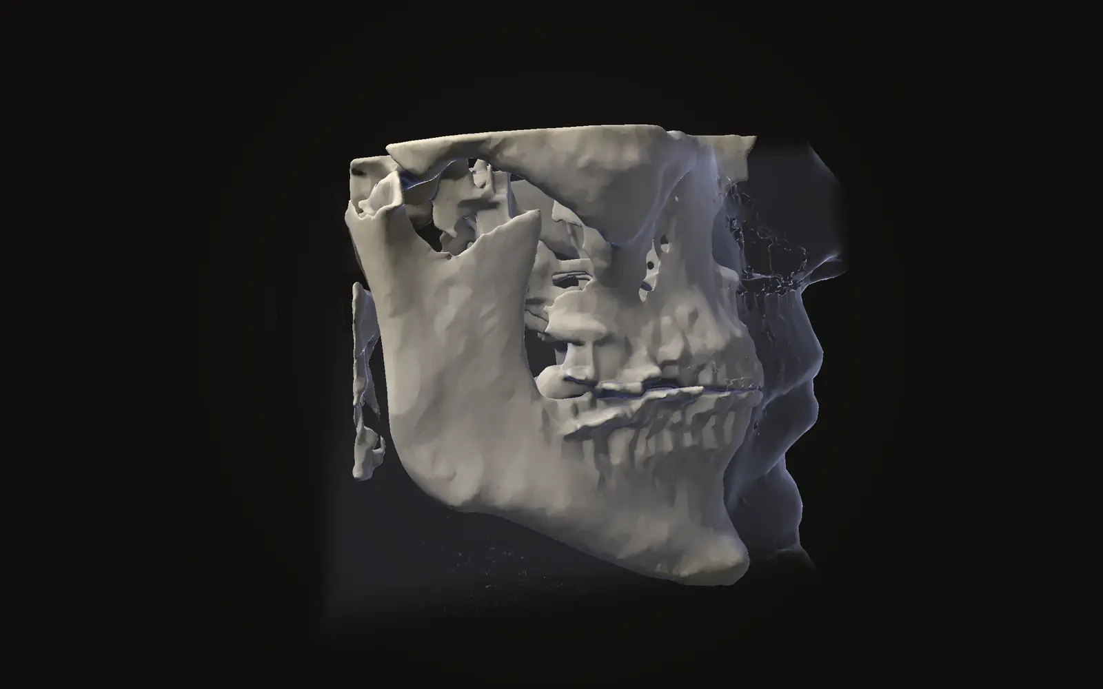
Jaw Viewer does not predict a face. It can open scans from before and after surgery, line them up on the skull base and show where the bone and skin changed. The comparison measures changes that have already happened. The cover image is the public sample scan in the viewer, not a patient outcome.
If the upper jaw moves 4 mm, does the lip move 4 mm?
No. One systematic review suggests about 0.6 mm of upper lip movement for every 1 mm of tooth movement in a Le Fort I advancement without an alar base cinch suture, and 0.9 mm with one. People vary widely around those averages.
Can a computer program predict exactly how my face will look?
No. The latest systematic review of 3D studies found no data that can be applied directly in daily practice. A prediction is an estimate with a range.
Does the lower lip follow the lower jaw one to one?
Not always. For advancement by sagittal split, a review found the lower lip moved about half as far as the lower incisors, while the chin soft tissue moved about as far as the chin bone.
Sources
- San Miguel Moragas J, Van Cauteren W, Mommaerts MY (2014). A systematic review on soft-to-hard tissue ratios in orthognathic surgery part I: maxillary repositioning osteotomy. J Craniomaxillofac Surg, 42: 1341-51.DOIPubMed
- Joss CU, Joss-Vassalli IM, Kiliaridis S, et al. (2010). Soft tissue profile changes after bilateral sagittal split osteotomy for mandibular advancement: a systematic review. J Oral Maxillofac Surg, 68: 1260-9.DOIPubMed
- Joss CU, Joss-Vassalli IM, Bergé SJ, et al. (2010). Soft tissue profile changes after bilateral sagittal split osteotomy for mandibular setback: a systematic review. J Oral Maxillofac Surg, 68: 2792-801.DOIPubMed
- San Miguel Moragas J, Oth O, Büttner M, et al. (2015). A systematic review on soft-to-hard tissue ratios in orthognathic surgery part II: Chin procedures. J Craniomaxillofac Surg, 43: 1530-40.DOIPubMed
- Ravelo V, Olate S, Bravo-Soto G, et al. (2025). Systematic review of soft-to-hard tissue ratios in orthognathic surgery: 3D analysis, update of scientific evidence. Int J Oral Maxillofac Surg, 54: 819-29.DOIPubMed
Terms used
- Soft-to-hard tissue ratio
- How far a point on the skin or lip moves for each millimetre the corresponding bone or tooth moves. A ratio of 0.6:1 means 0.6 mm of soft tissue for every 1 mm of bone.
- Le Fort I osteotomy
- A planned cut through the upper jaw above the teeth, so the whole tooth-bearing part can be moved and fixed in a new position.
- BSSO
- Bilateral sagittal split osteotomy. A planned cut along both sides of the lower jaw that lets the tooth-bearing part slide forward or back.
- Genioplasty
- An operation that cuts and repositions the chin bone.
This is general information, not medical advice. Jaw Viewer is not a medical device. Talk to your surgeon, orthodontist or radiologist about your own case.