Key points
- A 2020 review calls CBCT the standard three-dimensional X-ray technique in dental and jaw radiology; it has been in use there since the late 1990s.
- Bone, soft tissue and teeth are three tissue groups, and a 2011 review of 15 articles found that no single imaging method captures all three well. Combining images (image fusion) is how studies cover them.
- In a 2020 review of 12 studies, average differences between the digital model and the real outcome were under 2 mm in each direction for both jaws. Under 2 mm or 2 degrees was commonly judged acceptable.
- A 2014 review covered 7 trials with 149 patients. With a 2 mm mean-difference criterion, 125 of 133 patients in six of those articles had a successful outcome.
Orthognathic (jaw) surgery uses 3D images before and after the operation, and CBCT is the standard 3D X-ray technique in this field.[1] A single scan does not show everything the surgical team cares about, so studies combine it with other images and then check how closely the real result matches the digital model. This article describes what the published reviews report. It does not tell anyone what imaging they need.
Why CBCT?#
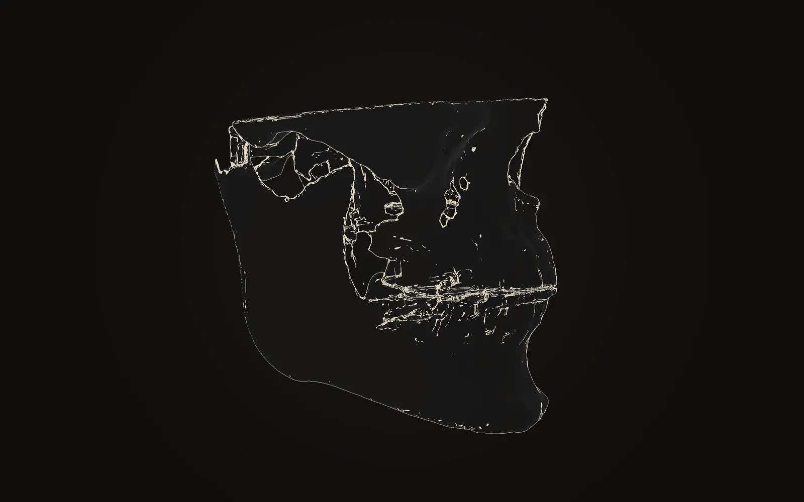
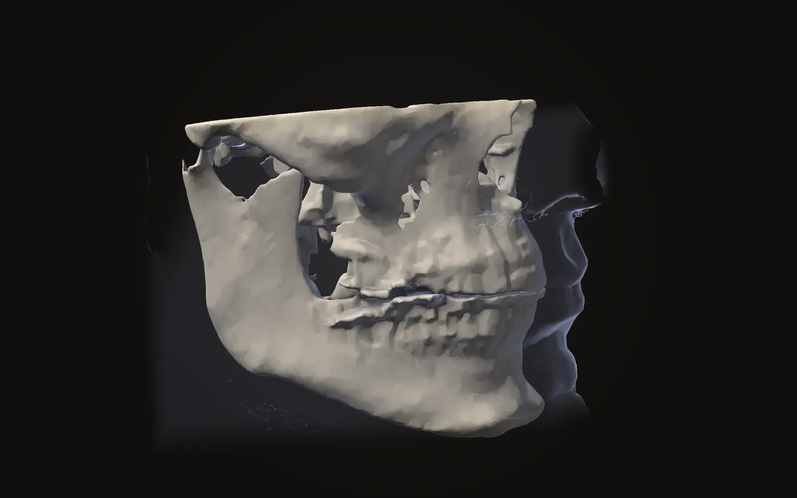
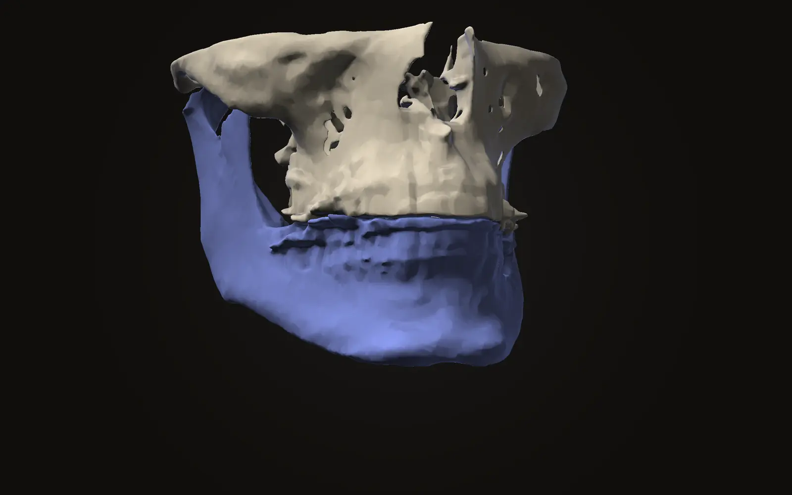
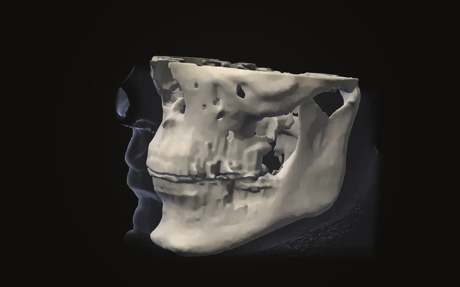
CBCT was first used in dental and jaw radiology at the end of the 1990s. It is now the standard three-dimensional X-ray technique in that field.[1] One turn around the head gives a stack of slices that can be shown from three directions or as a 3D model, which is why the same scan can serve both a close look at the jaw joint and an overview of the whole face. Our article on what a CBCT scan shows explains what those images are made of. The dose article covers the cost in radiation, and CBCT versus medical CT covers when a hospital CT is used instead.
On a jaw scan, bone, teeth and airway share one coordinate system. Distances and angles taken on one scan can be repeated on a later one, and two scans can be lined up and compared.
What does a scan not capture?#
Jaw surgery changes three things together: the soft tissue of the face, the bone underneath and the teeth. A 2011 systematic review of 15 articles calls these the triad and states that none of the craniofacial imaging techniques captures the whole triad with optimal quality.[2]
The review's answer was image fusion: merging two or more imaging techniques into one 3D virtual head that can show all three elements.[2] Its authors concluded that fused images are accurate for documentation, analysis and long-term follow-up.[2] It lists CT and MRI scanning, 3D photography and surface scanning as the technologies available for this.[2]
What are the images used for?#
The sources describe several uses for the images:
| Use | What the images provide |
|---|---|
| Documentation | A record of bone, teeth and soft tissue before the operation |
| Analysis | Distances, angles and volumes measured on the scan |
| Follow-up | A second scan, lined up with the first, to see what changed |
The first two are covered by the fusion review's list of documentation, analysis and follow-up.[2] The third needs the two scans to be registered, meaning lined up so the same anatomy sits in the same place. Where the lining-up goes wrong, the apparent change is partly an error of the method. We cover the size of that error in comparing two CBCT scans.
What do trials report about computer-made models of the operation?#
This is the one part of the topic that goes beyond viewing scans, and it is described here only as research. In the published trials, the team builds a digital model of the intended jaw movement before the operation. The studies then compare that model with a scan taken after the operation.[3]
A 2020 systematic review kept 12 clinical studies from 392 articles. It found an average translation of under 2 mm in the upper and lower jaw in all three directions. The reported rotation was under 2.75, 1.7 and 1.1 degrees (pitch, yaw, roll) for the upper jaw.[3] The review names CBCT combined with a scan of the dental casts as the most used imaging set-up, and notes that a difference under 2 mm or 2 degrees was commonly treated as acceptable.[3]
An earlier review set a similar bar. It found 7 trials with 149 patients and used a 2 mm mean difference as the success criterion in six articles. In those six, 125 of 133 patients had a successful outcome.[4] The authors could not pool the data, because the trials reported their results in different ways.[4]
under 2 mm
average translation, upper and lower jaw, 12 studies[3]
125 of 133 patients
successful at a 2 mm criterion, six articles[4]
These group averages do not describe a single operation. The studies define success differently, and both reviews call for more clinical trials.[3][4]
What does Jaw Viewer do here?#
Jaw Viewer opens a CBCT scan and lets you look at it: slices in three directions, an oblique slice, a 3D view with the skin faded so the bone and teeth show through, and a panoramic view. You can measure distances, angles, volumes and the airway, and compare two scans of the same person after they are lined up on the skull base. It does not build a model of an operation or test one, and it does not tell you whether a change is the intended result. See what the viewer does not do.
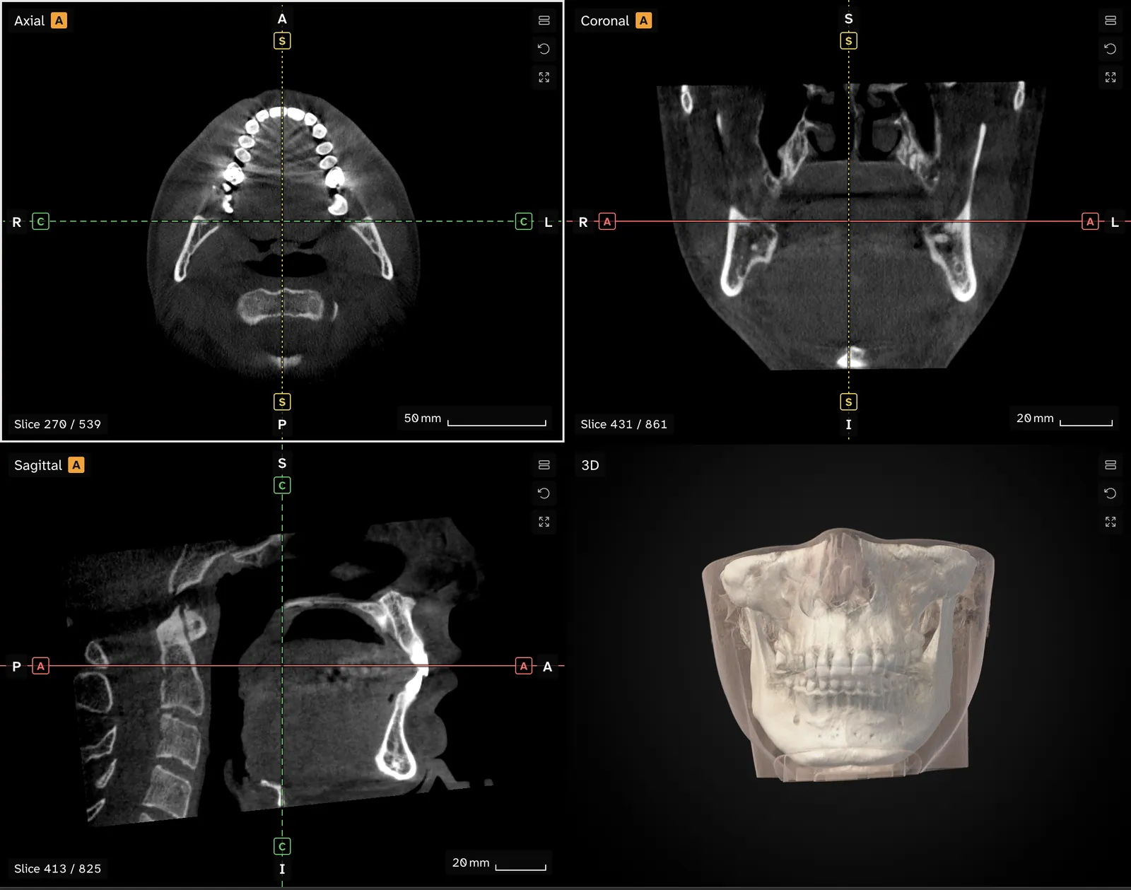
If you already have a scan, you can follow what a surgeon or radiologist shows you by looking at the same slices yourself. Your scan stays on your computer. See the privacy page.
What the sources leave open#
Both model-accuracy reviews rest on small numbers of clinical studies with different outcome measures.[3][4] The fusion review concerns how images are combined and tested, not the clinical result for patients.[2] None of them tells a reader which imaging is right for their own case. That decision belongs to the treating team. The sources here are from 2011 to 2020, and scanners and software have changed since.
Which scan is used for jaw surgery?
Reviews describe CBCT combined with a scan of the teeth or dental casts as the most used set-up for the digital models. CBCT is the standard 3D X-ray technique in dental and jaw radiology.
Does one scan show bone, soft tissue and teeth?
Not well. A 2011 review of 15 articles found that none of the imaging methods captures facial soft tissue, facial skeleton and teeth with optimal quality. Studies fuse several images instead.
How close is the real result to the digital model?
In a 2020 review of 12 studies, average differences were under 2 mm for both jaws. These are group averages from clinical studies, and they say nothing about an individual operation.
Sources
- Schulze RKW, Drage NA (2020). Cone-beam computed tomography and its applications in dental and maxillofacial radiology. Clin Radiol, 75: 647-57.DOIPubMed
- Plooij JM, Maal TJ, Haers P, et al. (2011). Digital three-dimensional image fusion processes for planning and evaluating orthodontics and orthognathic surgery. A systematic review. Int J Oral Maxillofac Surg, 40: 341-52. 15 articles on image fusion of two or more imaging techniques.DOIPubMed
- Alkhayer A, Piffkó J, Lippold C, et al. (2020). Accuracy of virtual planning in orthognathic surgery: a systematic review. Head Face Med, 16: 34. 12 clinical studies compared the digital model with the real outcome.DOIPubMed
- Stokbro K, Aagaard E, Torkov P, et al. (2014). Virtual planning in orthognathic surgery. Int J Oral Maxillofac Surg, 43: 957-65. 7 clinical trials, 149 patients. No meta-analysis was possible.DOIPubMed
Terms used
- CBCT
- Cone beam computed tomography. A scanner that rotates once around the head and records a cone-shaped X-ray beam, common in dentistry and jaw surgery.
This is general information, not medical advice. Jaw Viewer is not a medical device. Talk to your surgeon, orthodontist or radiologist about your own case.