Jaw surgery and orthodontics
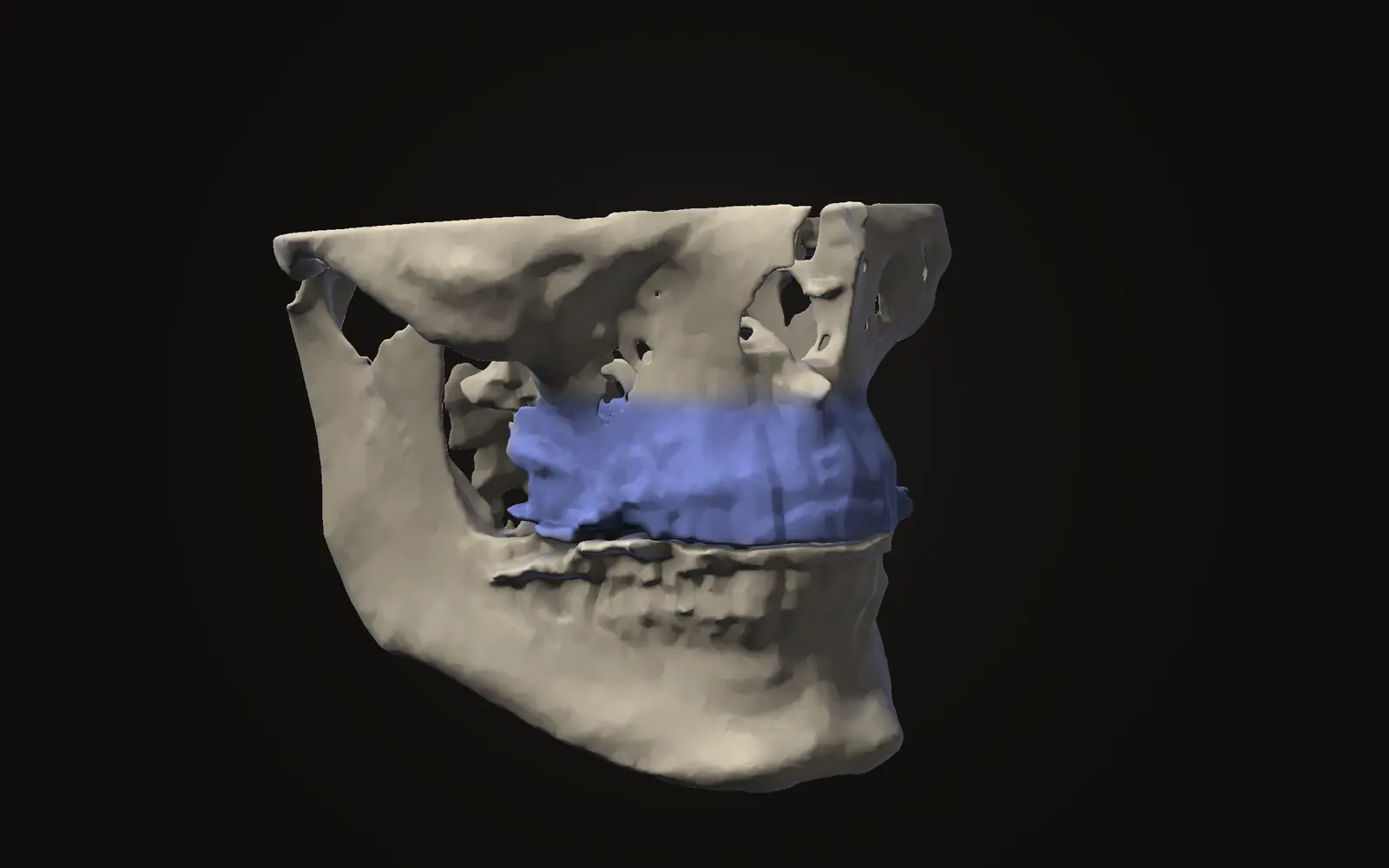
Le Fort I osteotomy: the upper jaw operation explained
A Le Fort I osteotomy frees the upper jaw to move in all three planes. What reviews report on stability and complications, and how weak that evidence is.
What jaw scans show, what jaw surgery changes, and how to read both. Every claim links to its source.
Jaw surgery and orthodontics
A Le Fort I osteotomy frees the upper jaw to move in all three planes. What reviews report on stability and complications, and how weak that evidence is.
Measurements explained
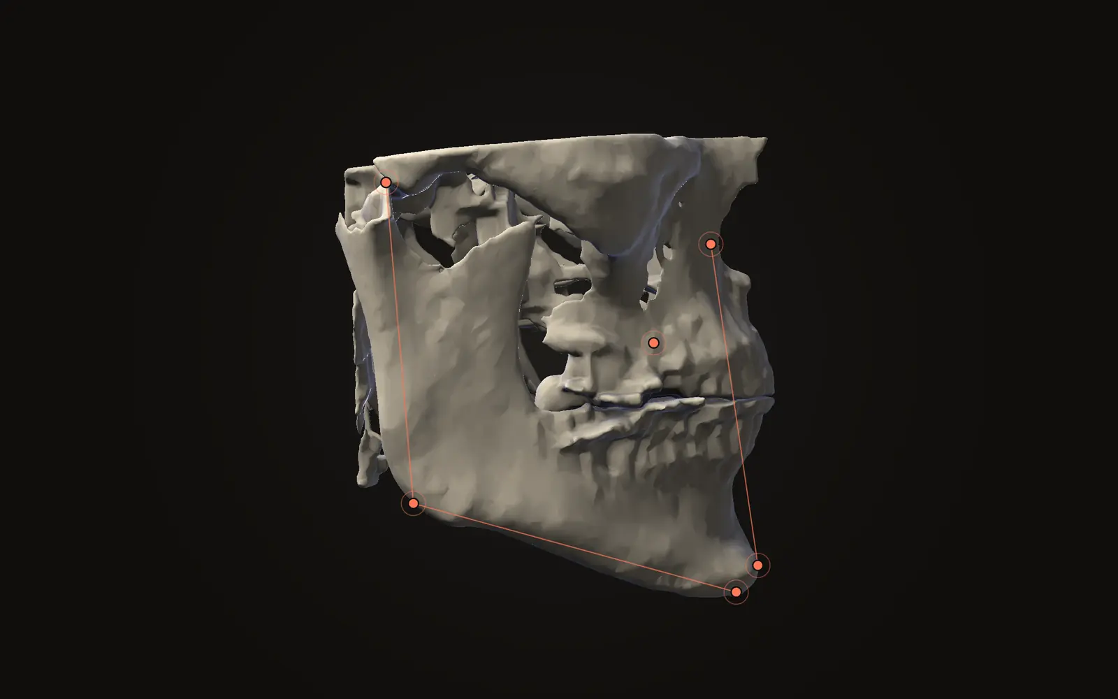
Cephalometric angles are built from hand-placed points that never land twice in the same spot. What reviews report about which landmarks hold steady.
Using Jaw Viewer
How to check whether a free scan viewer uploads your CBCT or DICOM files, using the Network panel in your browser, and what that check cannot show.
Measurements explained
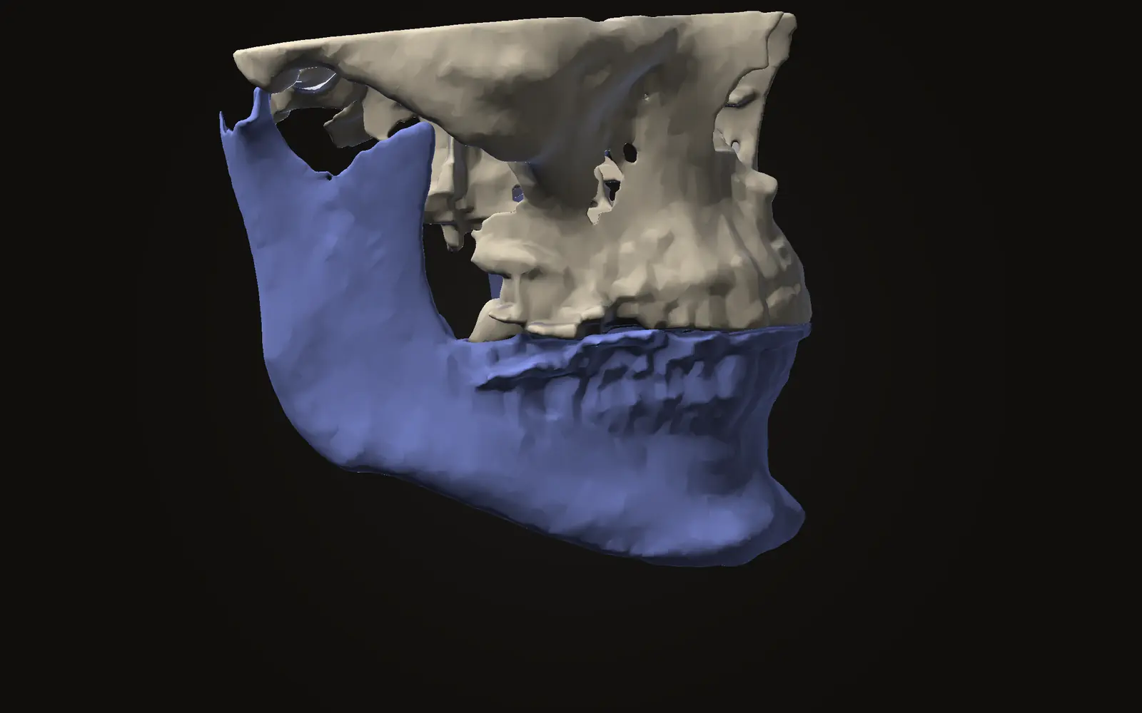
Where the inferior alveolar nerve canal runs in the lower jaw, how it shows on a CBCT scan, and how much it varies between people, with review figures.
Measurements explained
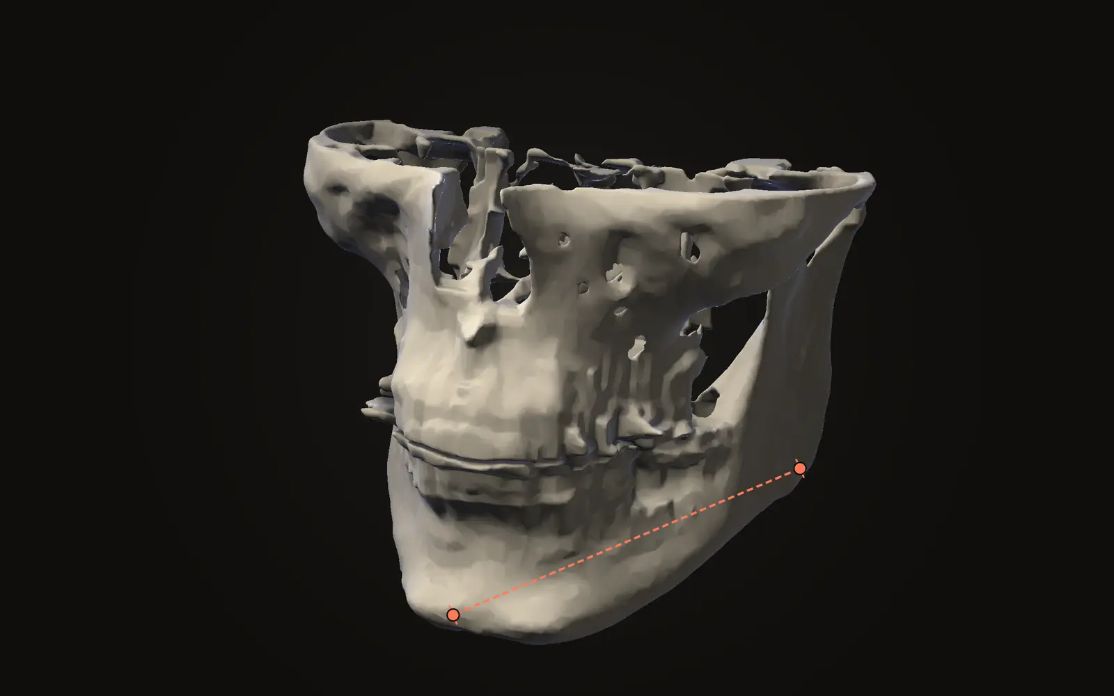
Two points make a distance, three an angle, a closed outline an area. How each measurement works on a scan, in which view to place it, and what to check.
Measurements explained
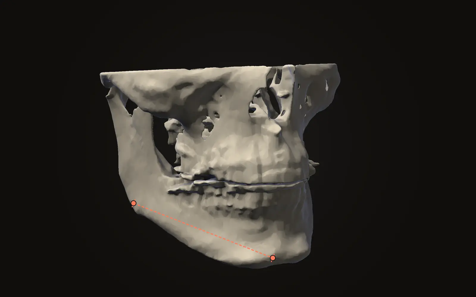
Reviews of CBCT linear measurements report a wide error range, over and under. What the studies tested, what voxel size does and what stays open.
Measurements explained
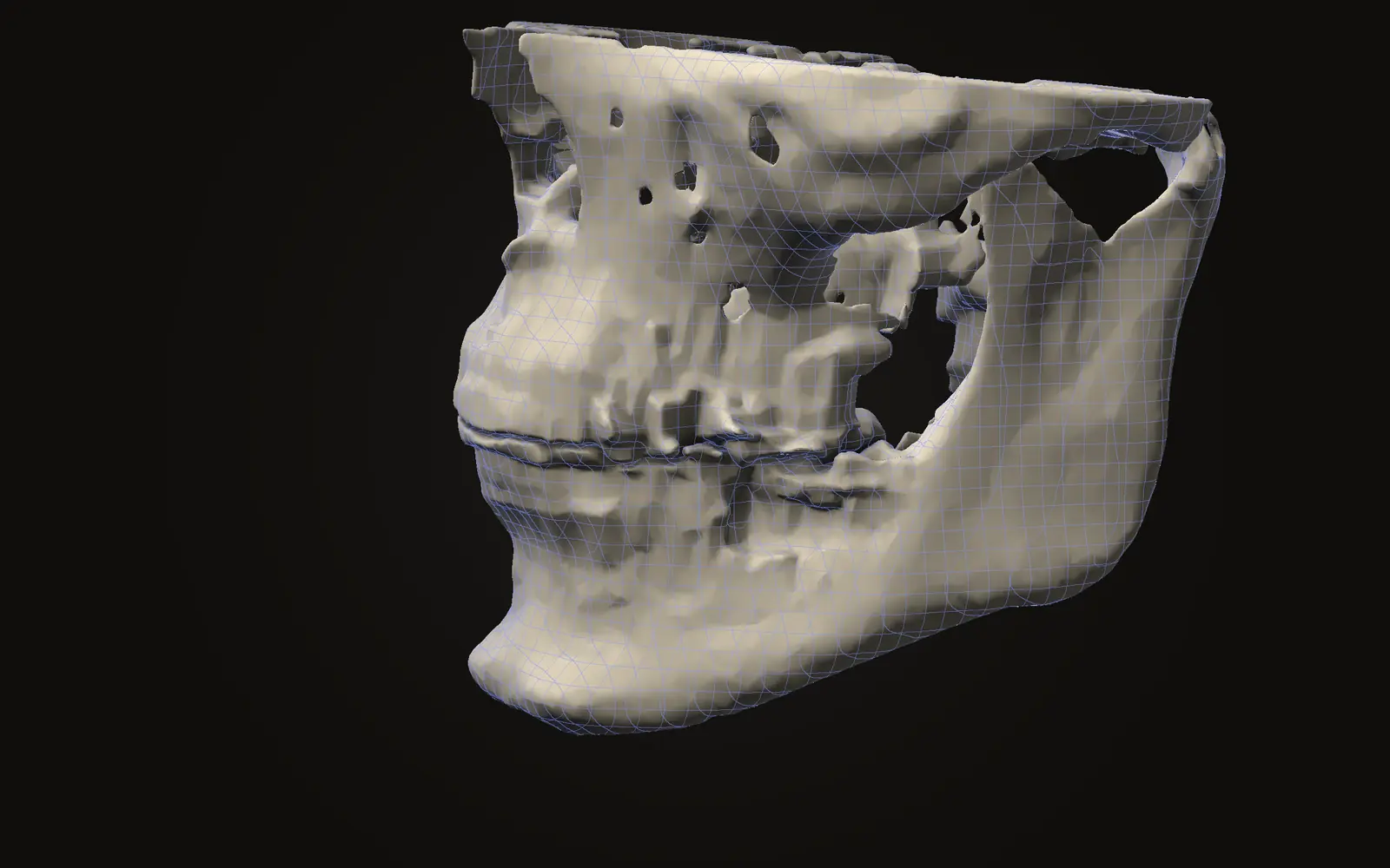
CBCT grey values are not calibrated Hounsfield units. What causes the gap, what phantom and calibration studies found, and what a viewer's number means.
Jaw surgery and orthodontics
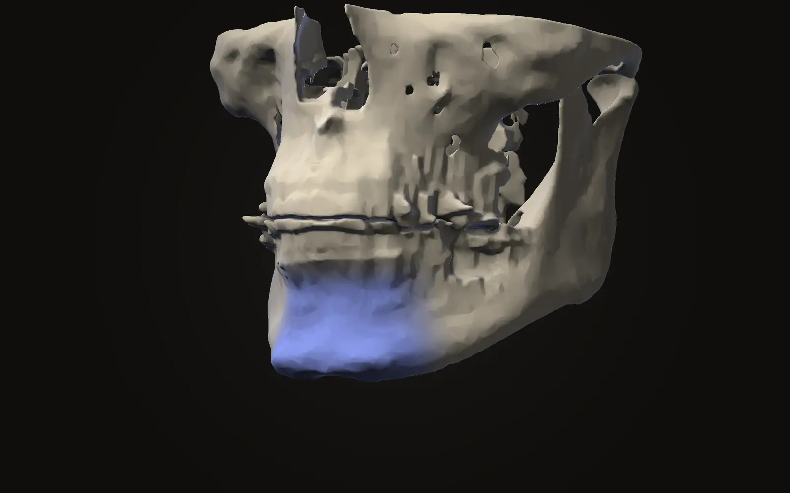
Genioplasty cuts and repositions the chin bone. What reviews report on how the skin follows the bone and on relapse, and how thin the evidence still is.
Using Jaw Viewer
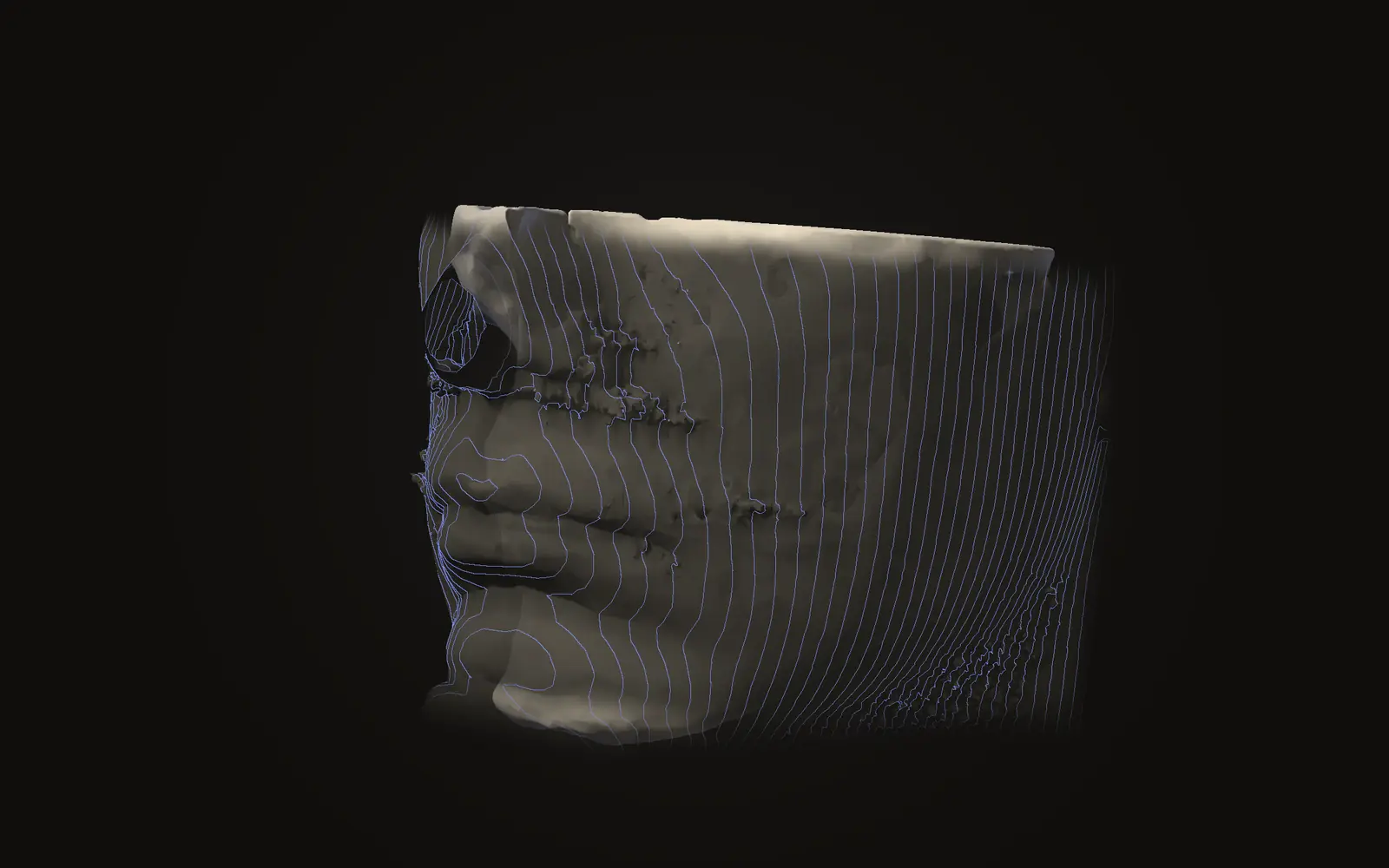
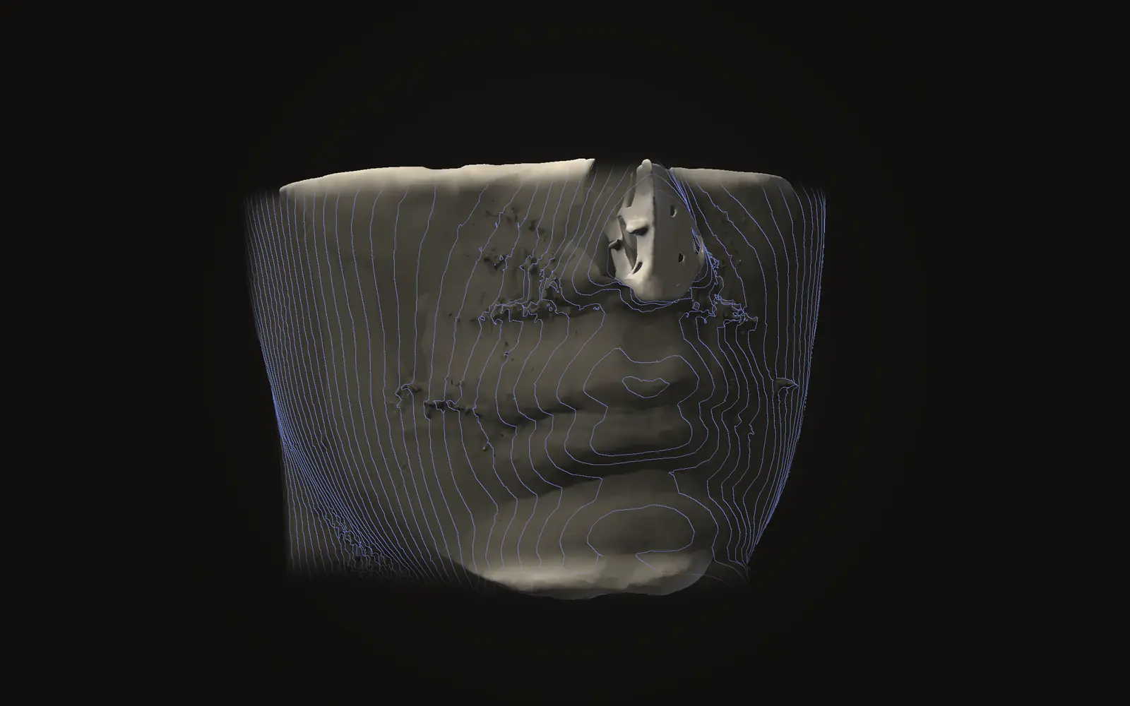
A head scan can be drawn as a 3D face, and software has matched such faces to photographs. Removing the name from the files does not remove the face.
Measurements explained
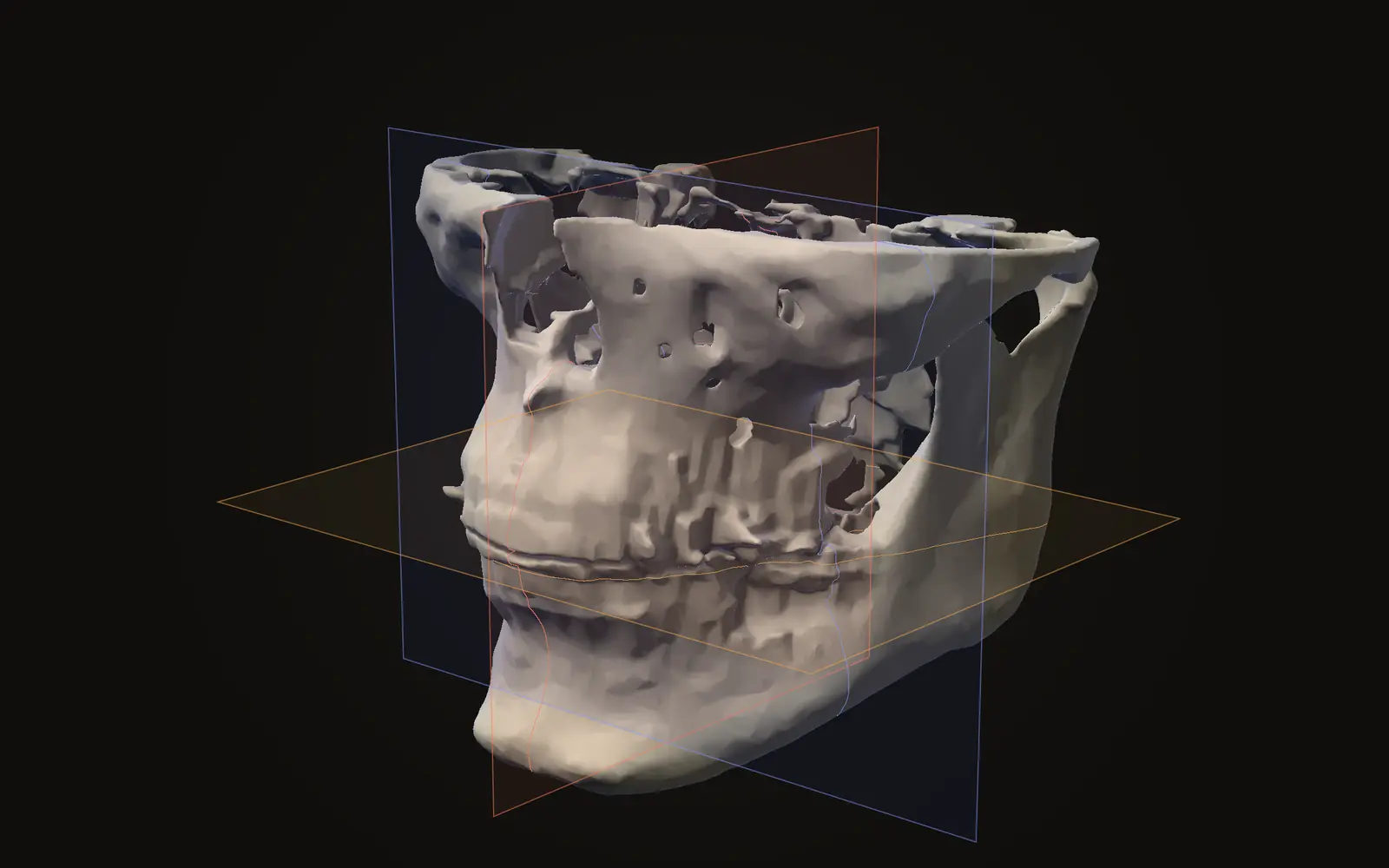
Registration lines up two CBCT scans before you compare them. Research reports 0.2 to 0.45 mm of leftover gap, and the reference region matters.
Jaw surgery and orthodontics
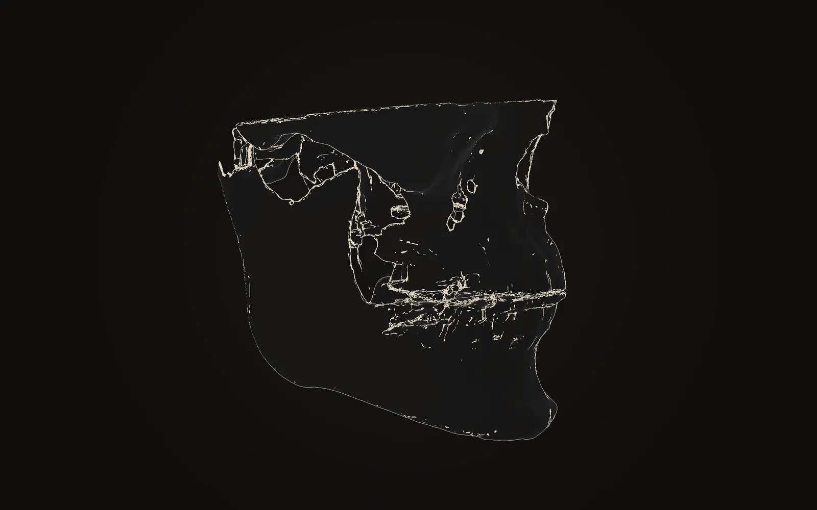
Two CBCT scans of one person are lined up on the skull base, which jaw surgery does not move, so that changes elsewhere can be shown as colour.
Understanding your scan
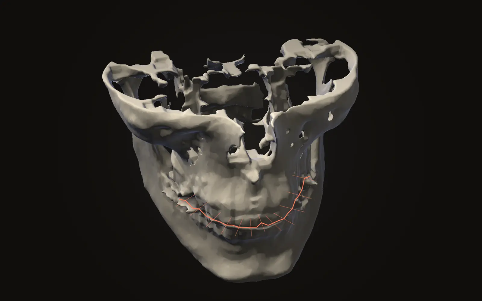
A panoramic X-ray is one flat image, a CBCT scan is a 3D stack of slices. What each shows, what one study found about accuracy, and how the doses compare.
Articles are written from peer-reviewed papers and guidelines, and checked again at least once a year. Nothing here is medical advice.